Category: Visual Diagnosis
Posted: 1/13/2014 by Haney Mallemat, MD
Click here to contact Haney Mallemat, MD
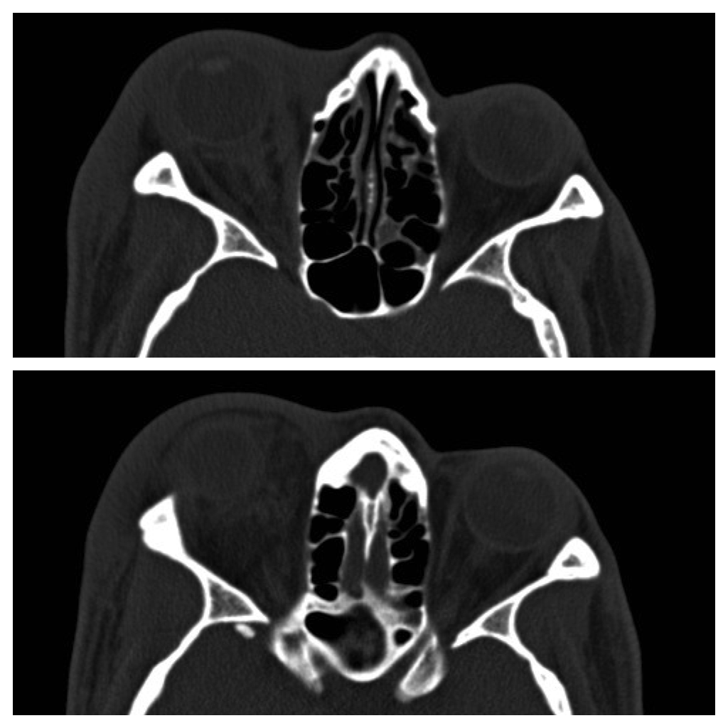
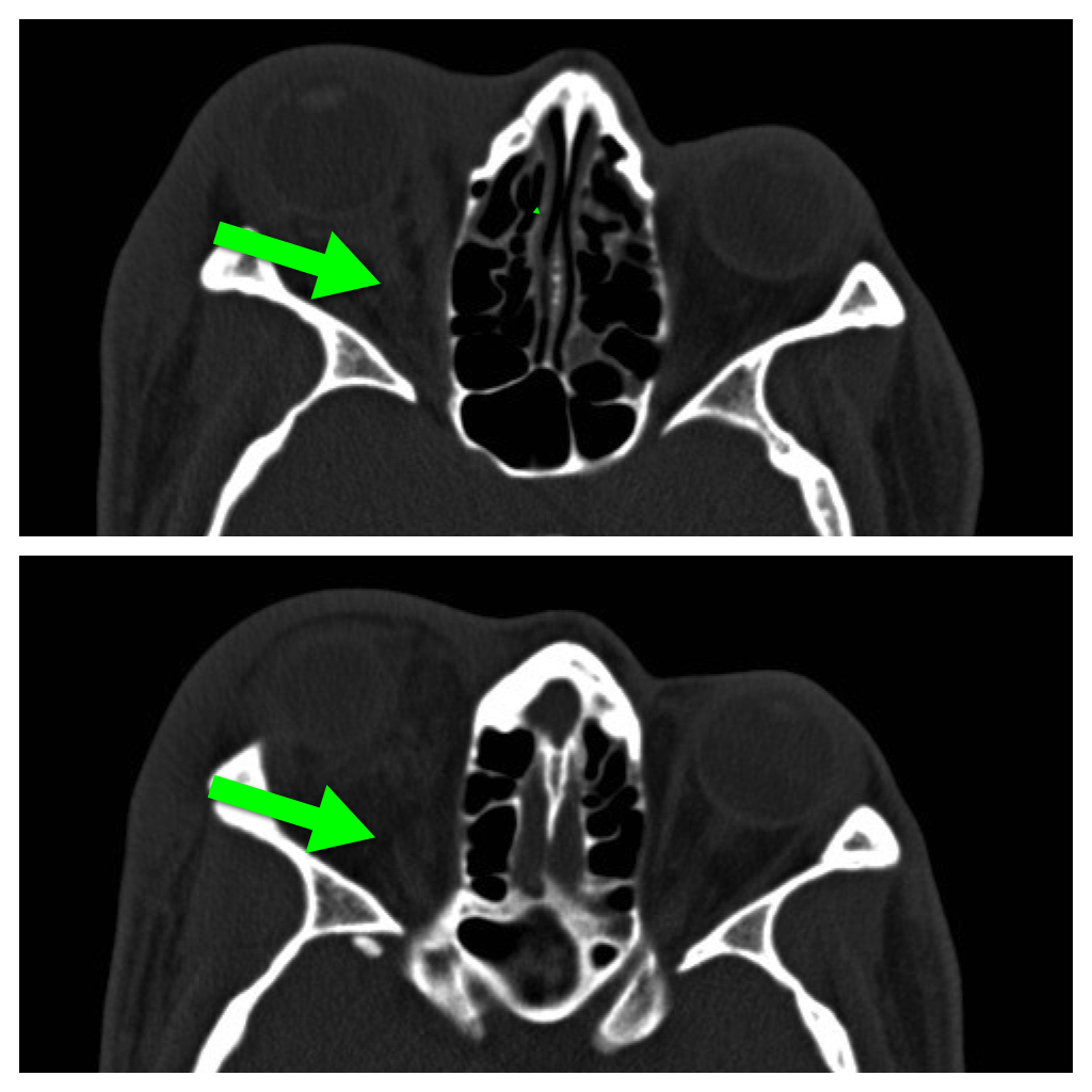
42 year-old male s/p assault complains of right sided facial pain, swelling, and decreased vision. Physical exam reveals subconjunctival hemorrhage, proptosis, afferent pupillary defect, and a firm globe. What's the diagnosis and what's the emergent treatment?
Follow me on Twitter (@criticalcarenow) or Google+ (+criticalcarenow)
Category: Visual Diagnosis
Posted: 1/6/2014 by Haney Mallemat, MD
Click here to contact Haney Mallemat, MD
37 year-old male presents after sustaining a burn from a pot of boiling water. He states that his skin started to blister a few hours after and it’s quite painful. What type of burn does he likely have?
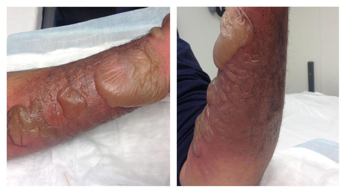
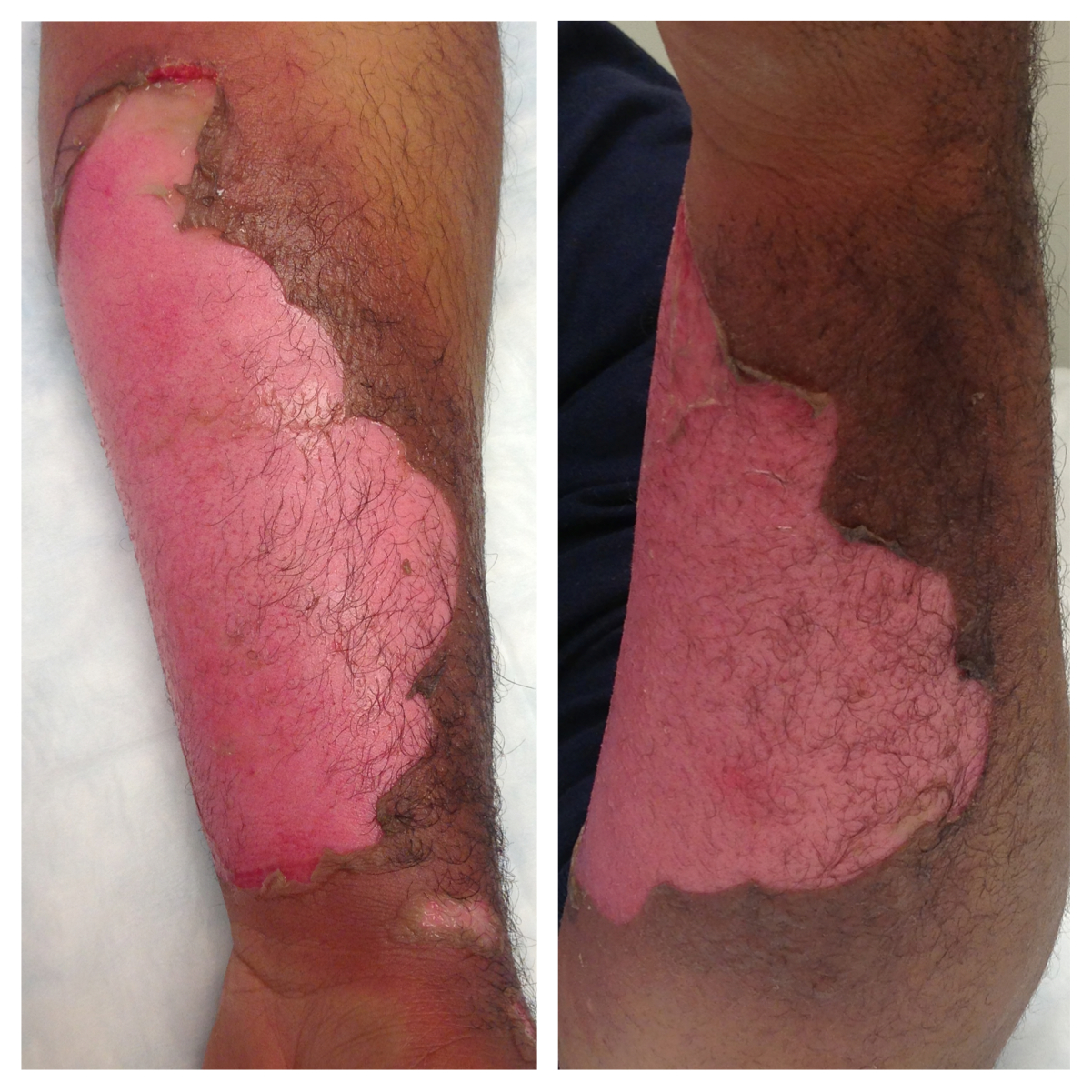
A non-circumferential, superficial partial-thickness burn; it was treated with Silvadene (silver sulfadiazine)
Burn Classification:
Follow me on Twitter (@criticalcarenow) or Google+ (+criticalcarenow)
Tintinalli, Judith E. (2010). Emergency Medicine: A Comprehensive Study Guide (Emergency Medicine). New York: McGraw-Hill Companies. pp. 1374–1386.
Category: Visual Diagnosis
Posted: 12/30/2013 by Haney Mallemat, MD
Click here to contact Haney Mallemat, MD
68 year-old male presents with weakness after surgical repair of his abdominal aorta. What’s the diagnosis and name at least one eponym for the signs displayed (there are five total)?
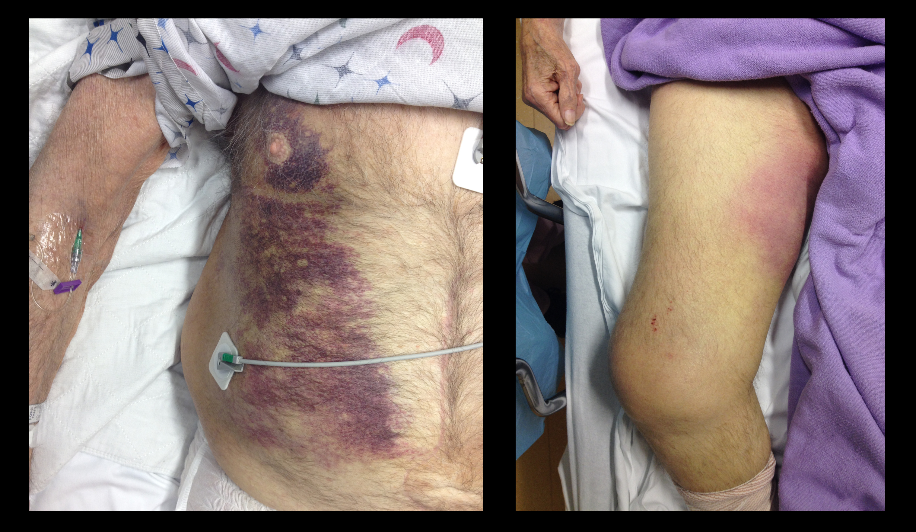
Grey-Turner and Fox's sign; these signs indicate retroperitoneal hemorrhage.
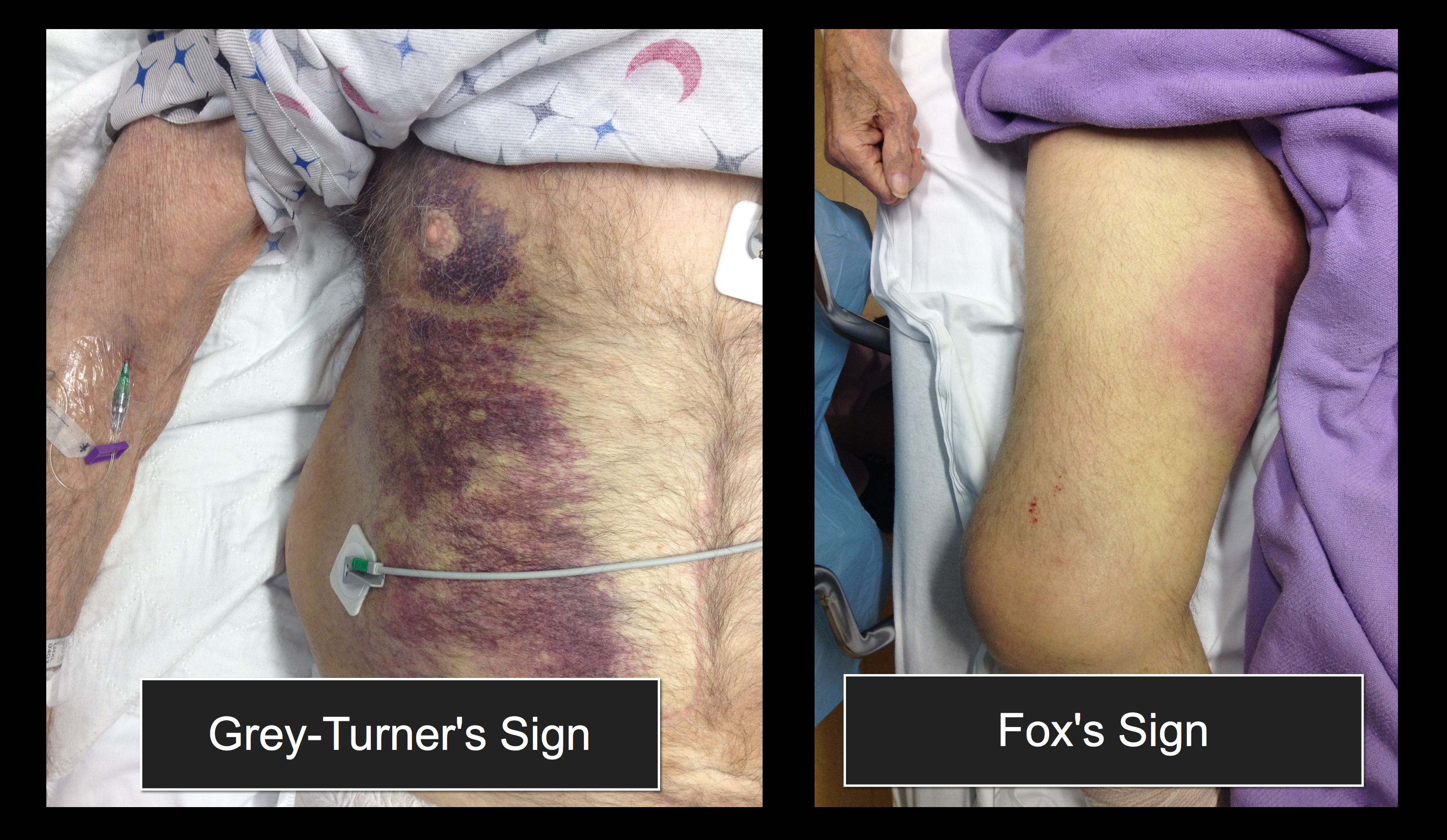
There are five signs suggesting retroperitoneal bleeding. They generally appear 24 hours after bleeding starts, occurring when blood extravasates along ligamentous connections between the retroperitoneal space and skin surface.
1. Grey-Turner Sign: Named for the surgeon who identified it in 1920; a bluish hematoma across the lateral abdominal wall when blood from the pararenal space leaks along the quadratus lumborum.
2. Fox’s Sign: Described by Dr. Fox in 1966 in two patients, (ruptured AAA and pancreatitis); ecchymosis over anteromedial thigh secondary to blood seeping along the fascia of psoas and iliacus.
3. Cullen’s Sign: Dr. Cullen (gynecologist), described this in 1918 as a sign of a ruptured ectopic pregnancy; bruise around the umbilicus from retroperitoneal blood tracking along falciform ligament.
4. Bryant’s Sign: Dr. Bryant initially described this sign; ecchymosis of the scrotum from blood tracking down the spermatic cord.
5. Stabler’s sign: Ecchymosis over the inguinal ligament
Follow me on Twitter (@criticalcarenow) or Google+ (+criticalcarenow)
Category: Visual Diagnosis
Posted: 12/23/2013 by Haney Mallemat, MD
Click here to contact Haney Mallemat, MD
Patient with a history of osteogenesis imperfecta presents with right lateral chest pain following a sneeze. The ultrasound of his chest is shown (hint: arrow points to a rib). What's the diagnosis?
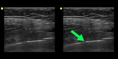
Rib fracture
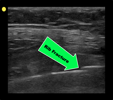
Turk F. et al. Evaluation by ultrasound of traumatic rib fractures missed by radiography. Emerg Radiol. 2010 Nov;17(6):473-7. doi: 10.1007/s10140-010-0892-9
Follow me on Twitter (@criticalcarenow) or Google+ (+criticalcarenow)
Category: Visual Diagnosis
Posted: 12/16/2013 by Haney Mallemat, MD
Click here to contact Haney Mallemat, MD
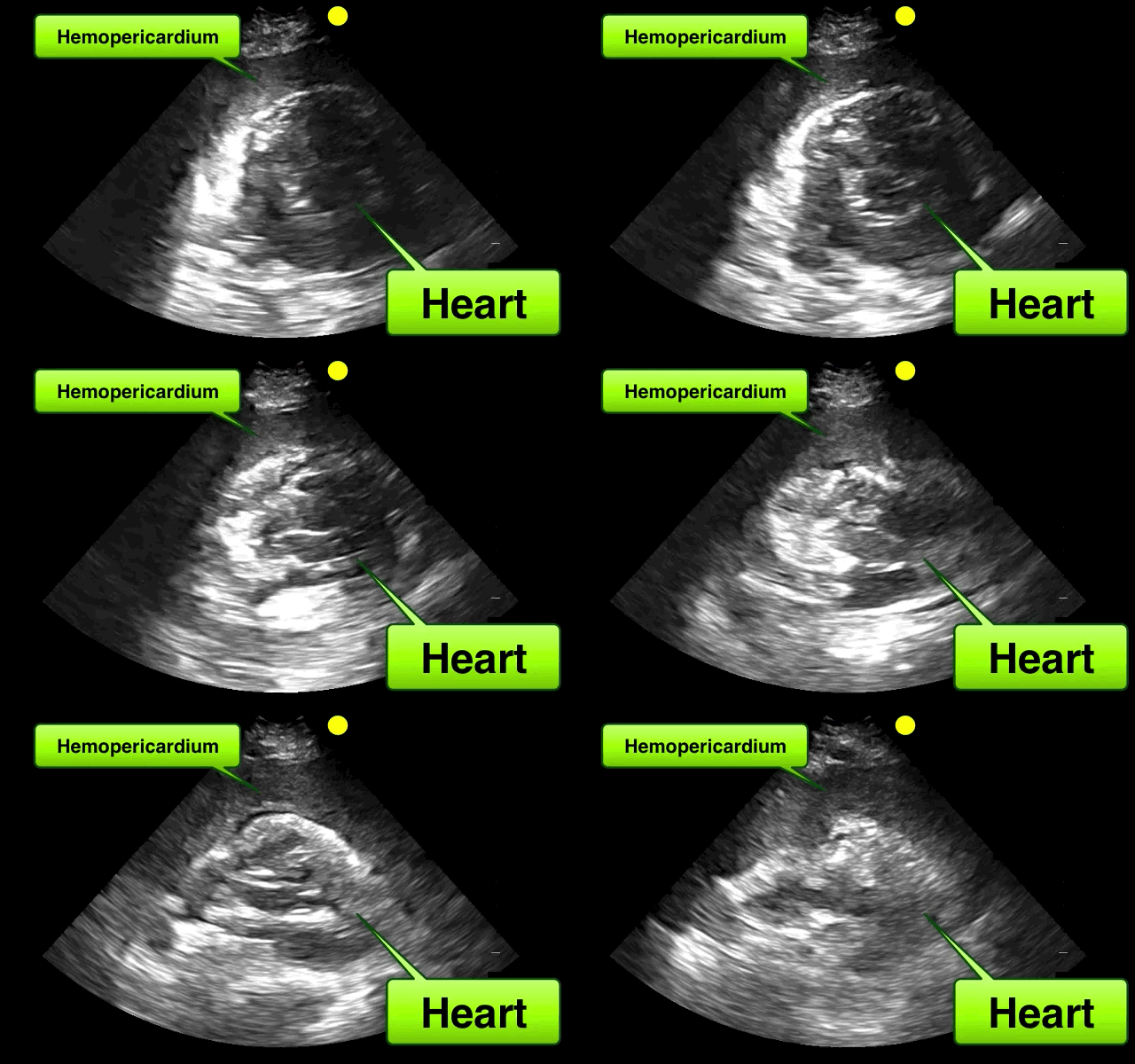
46 year-old female found unresponsive at a party. EMS transports the patient in cardiac arrest. A parasternal-long axis view of the heart is obtained during the pulse check. What's the diagnosis?
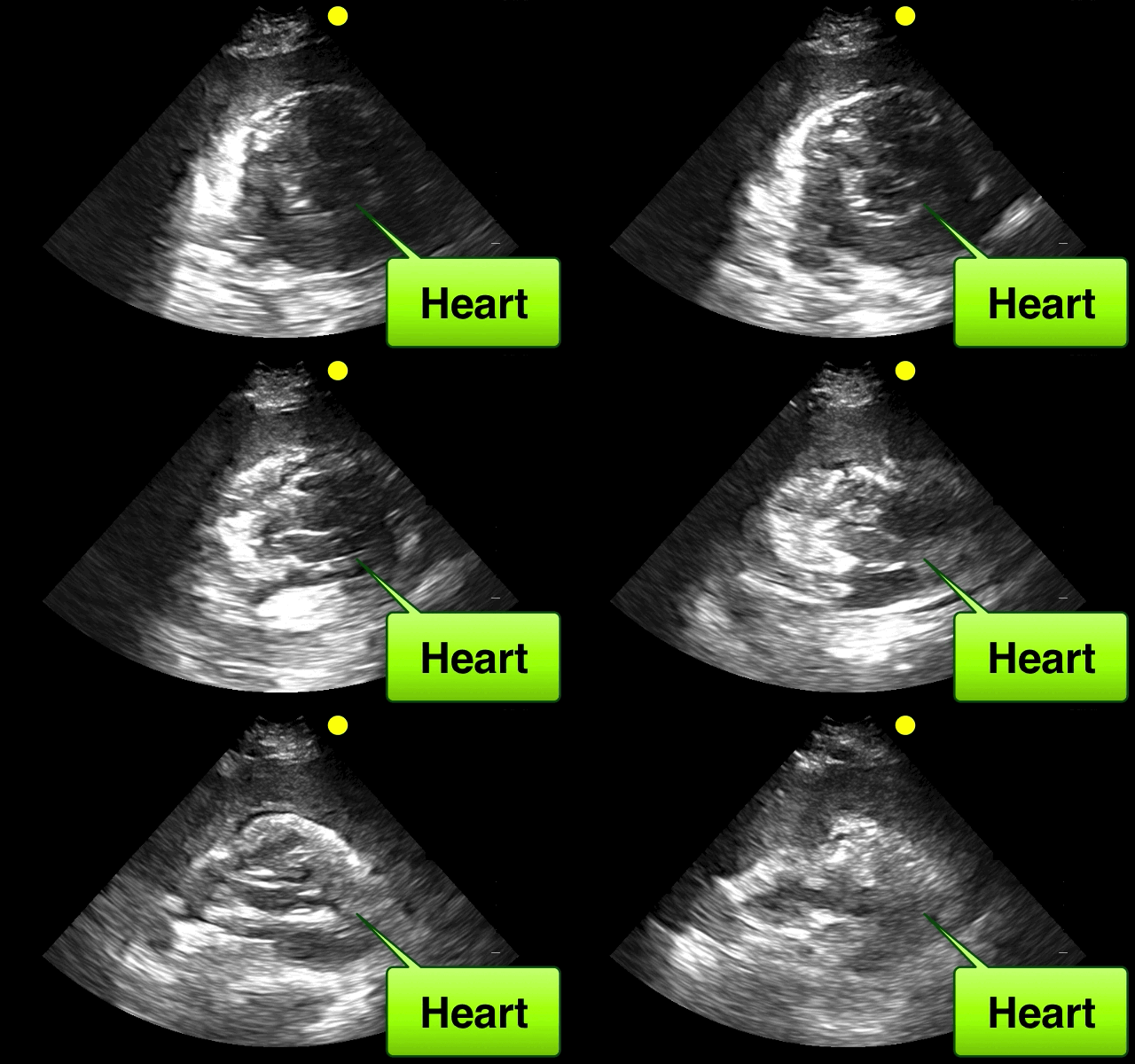
Hemopericardium
The heterogeneous appearance of the pericardial fluid indicates that is likely a complex pericardial effusion; the fluid could be blood, pus, or a malignant effusion.
Differential diagnosis of hemopericardium includes:
Based on this initial ECHO, a pericardiocentesis was performed and blood was aspirated.
Follow me on Twitter (@criticalcarenow) or Google+ (+criticalcarenow)
Category: Visual Diagnosis
Posted: 12/9/2013 by Haney Mallemat, MD
Click here to contact Haney Mallemat, MD
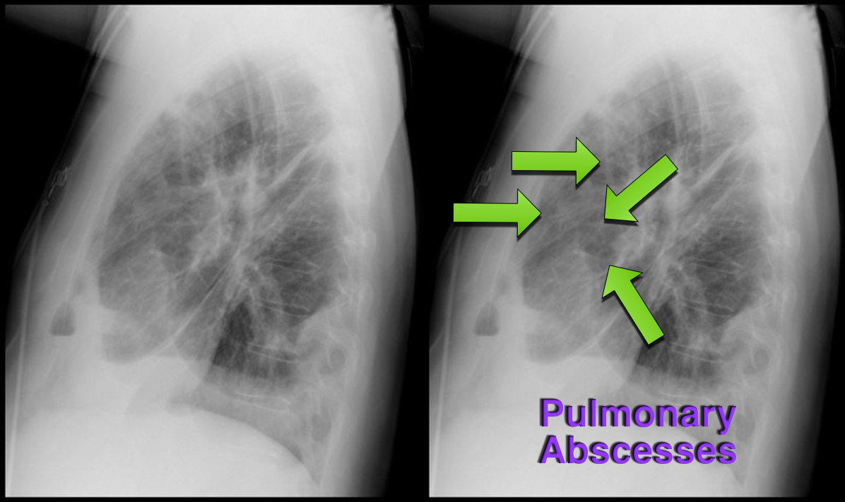
37 year-old male presents with cough and a fever. What's the diagnosis and name three risk factors assiciated with disease?
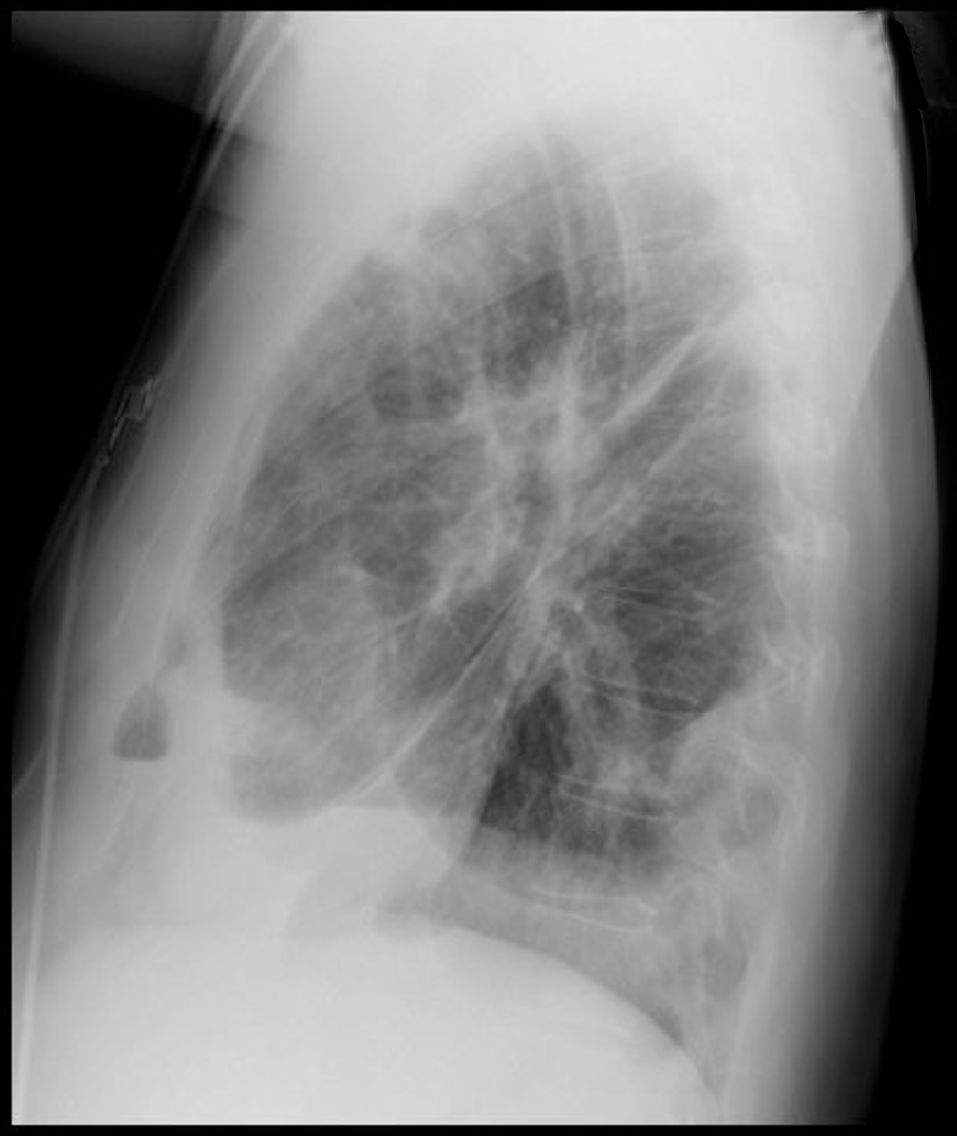
Answer: Lung Abscesses
Lung Abscess
Follow me on Twitter (@criticalcarenow) or Google+ (+criticalcarenow)
Category: Visual Diagnosis
Posted: 12/2/2013 by Haney Mallemat, MD
Click here to contact Haney Mallemat, MD
Which view of the heart is this and can you name the structures from A-G?
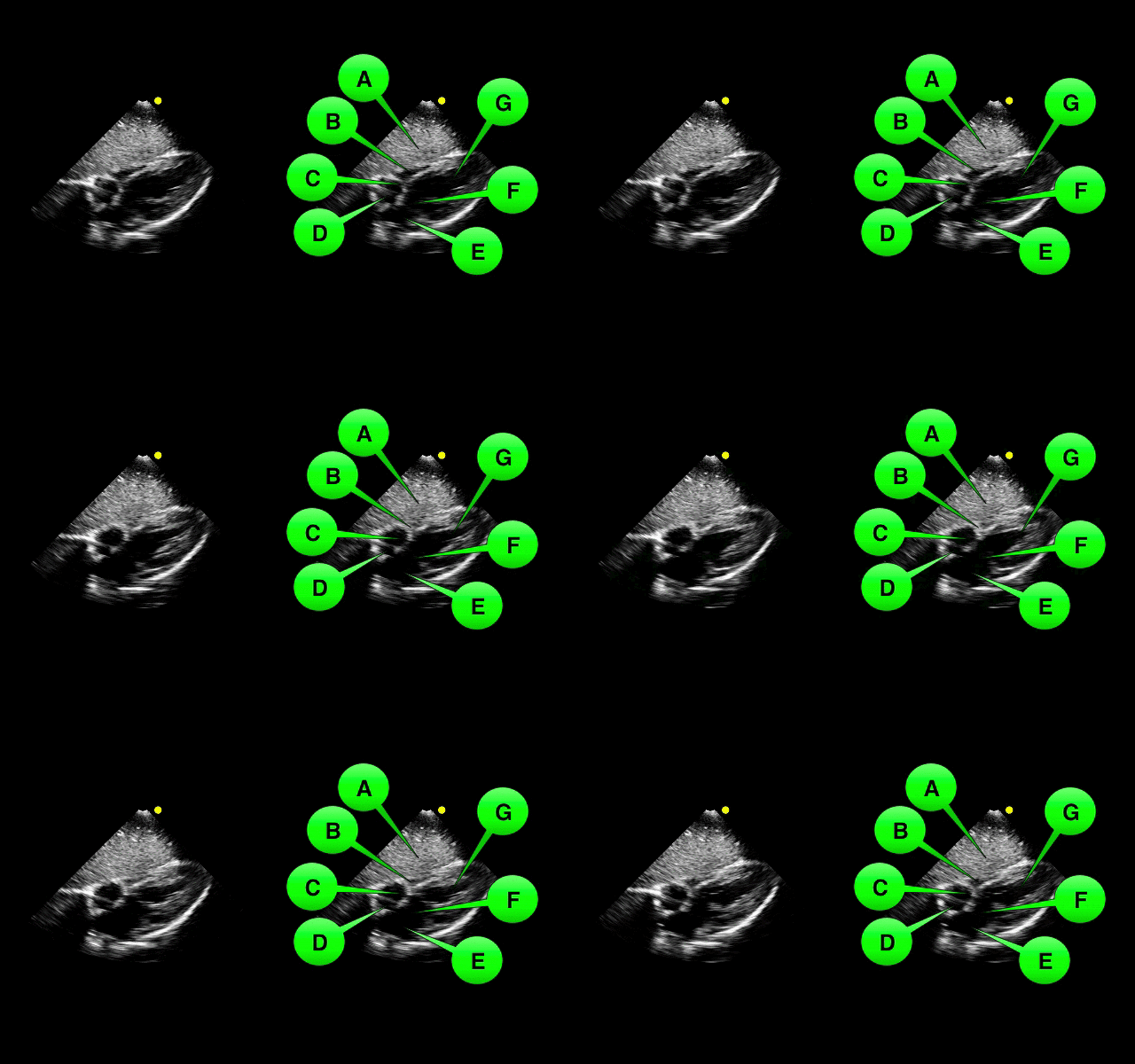
1. Subcostal or Subxiphoid view; this view is obtained by placing the probe under the ribs with the patient supine. The liver is used as an acoutic window to image the heart.
2. Name the items labeled A-G:

Follow me on Twitter (@criticalcarenow) or Google+ (+criticalcarenow)
Category: Visual Diagnosis
Posted: 11/25/2013 by Haney Mallemat, MD
Click here to contact Haney Mallemat, MD
What view of the heart is this and can you name everything from A-G?
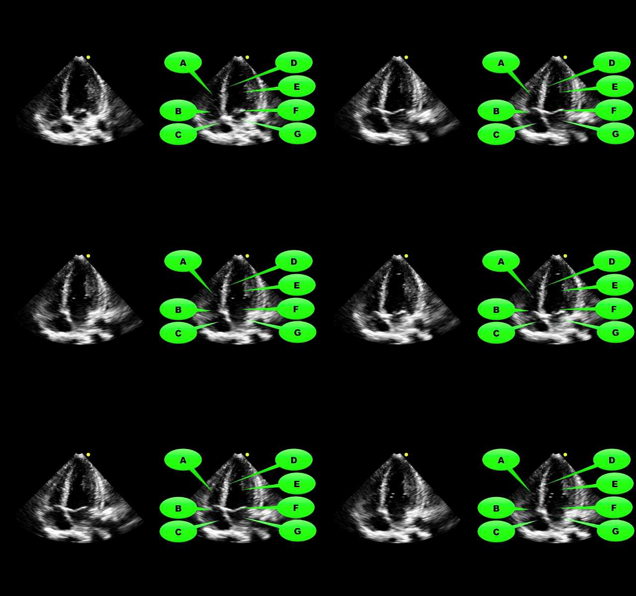
1. Apical-four chamber view; this view is obtained by placing the probe in the 4-5th intercostal space at the anterior axillary-line. The patient can be placed in left lateral decubitus to improve imaging.
2. Name the items labeled A-G:

Follow me on Twitter (@criticalcarenow) or Google+ (+criticalcarenow)
Category: Visual Diagnosis
Posted: 11/18/2013 by Haney Mallemat, MD
Click here to contact Haney Mallemat, MD
48 year-old presents after falling 15 feet following a “misunderstanding” with police. What's the diagnosis? ...and for a bonus question, why is this called a “Lover’s Fracture”?
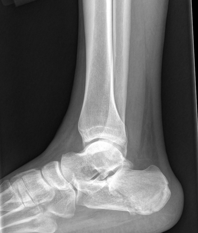
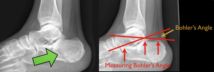
Calcaneus fracture
Answer to Bonus Question: Historically called a “Lover’s Fracture” for “lovers” jumping out of bedroom windows (to evade suspicious spouses) who then land directly on their feet.
Calcaneus fractures
Follow me on Twitter (@criticalcarenow) or Google+ (+criticalcarenow)
Category: Visual Diagnosis
Posted: 11/11/2013 by Haney Mallemat, MD
Click here to contact Haney Mallemat, MD
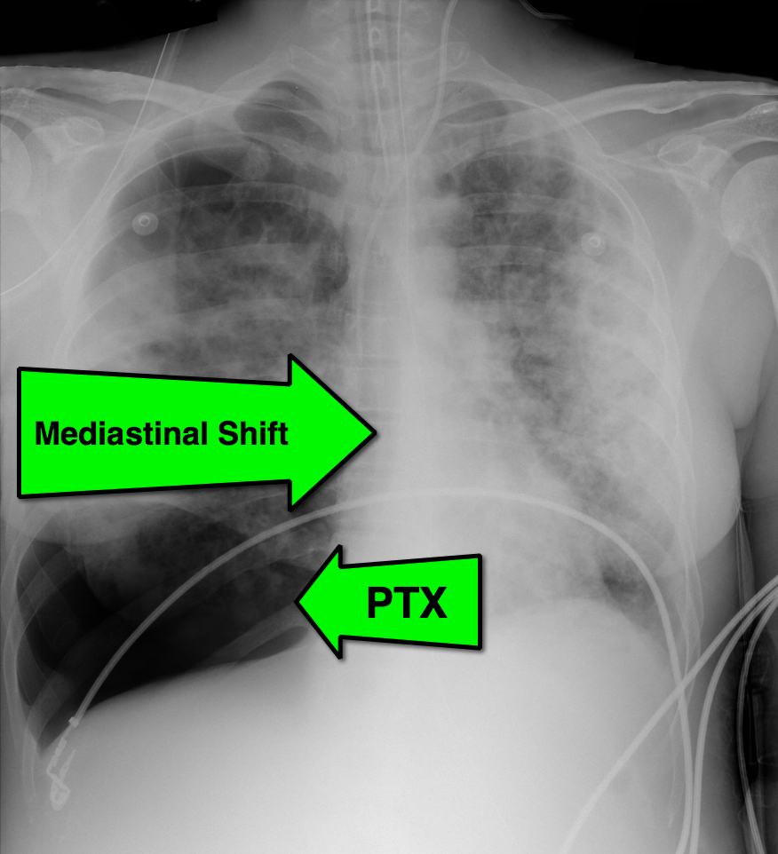
28 year-old cachectic female presents in respiratory distress and is immediately intubated on arrival to Emergency Department. What's the diagnosis and what are some potential etiologies?
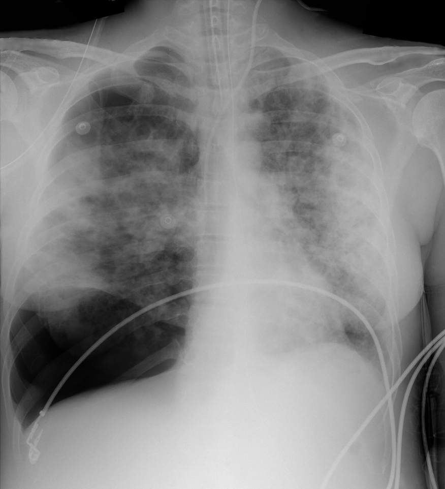
Pneumothorax with mediastinal shift
Differential Diagnosis
The patient in this case had undiagnosed HIV/AIDS and presented with PTX secondary to PJP. The lifetime risk of PTX with HIV is 6% and 85% of those cases are secondary to PJP.
Follow me on Twitter (@criticalcarenow) or Google+ (+criticalcarenow)
Category: Visual Diagnosis
Posted: 11/4/2013 by Haney Mallemat, MD
Click here to contact Haney Mallemat, MD
This week's visual pearl reviews the structures of the heart when being viewed in a parasternal long-axis view. What do the labels correspond to in the clip below (note: "E" and "F" are valves) and do you see any obvious abnormalities?
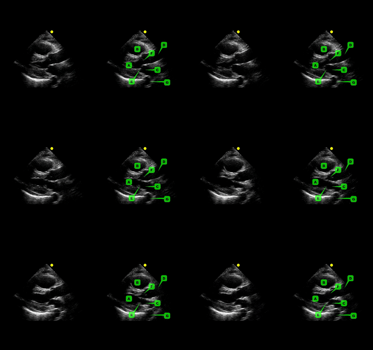
The parasternal long-axis is obtained by scanning to the left (patient's left) of the sternum through the 2nd-5th intercostal space. Click here for a tutorial on the technique.
Answer to Bonus Question: Dilation of the RVOT
Follow me on Twitter (@criticalcarenow) or Google+ (+criticalcarenow)
Category: Visual Diagnosis
Posted: 10/27/2013 by Haney Mallemat, MD
(Updated: 10/28/2013)
Click here to contact Haney Mallemat, MD
15 year-old right-hand dominant male received a direct blow to the right arm with a hockey stick. What’s the diagnosis?
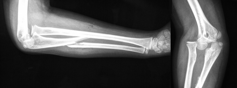
Monteggia Fracture
Follow me on Twitter (@criticalcarenow) or Google+ (+criticalcarenow)
Category: Visual Diagnosis
Posted: 10/20/2013 by Haney Mallemat, MD
(Updated: 12/5/2023)
Click here to contact Haney Mallemat, MD
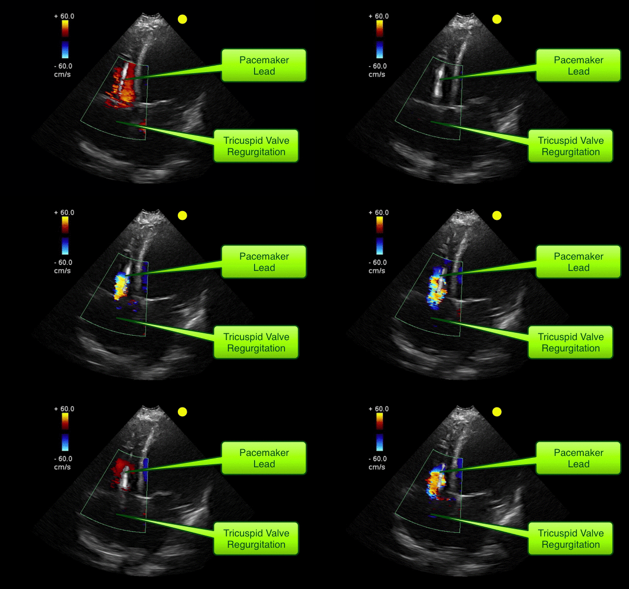
55 year-old male presents with chest pain. You take a look at his cardiac function with ultrasound and here's the patient's apical four-chamber view. What's in his right ventricle and why would it be there?
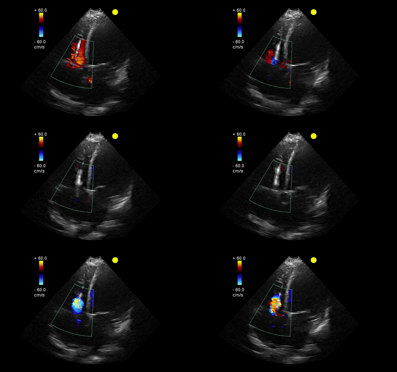
AICD
Follow me on Twitter (@criticalcarenow) or Google+ (+criticalcarenow)
Category: Visual Diagnosis
Posted: 10/14/2013 by Haney Mallemat, MD
Click here to contact Haney Mallemat, MD
A 23 year-old male presents with the rash below. He originally presented to his primary care doctor for a sore throat and was given a prescription for a medication; this rash subsequently broke out. What's the diagnosis and which medication did he receive?

Rash secondary to Epstein-Barr pharyngitis treated with amoxicillin
Luzuriaga, K., Sullivan, J. Infectious Mononucleosis. N Engl J Med 2010; 362:1993-2000
Follow me on Twitter (@criticalcarenow) or Google+ (+criticalcarenow)
Category: Visual Diagnosis
Posted: 10/7/2013 by Haney Mallemat, MD
Click here to contact Haney Mallemat, MD
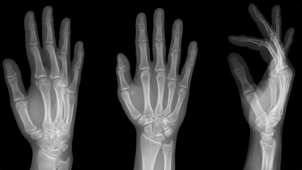
25 year-old female struck in the left hand by a football. Presents with pain, visible deformity, and the Xray below. What are the next step(s) in management?
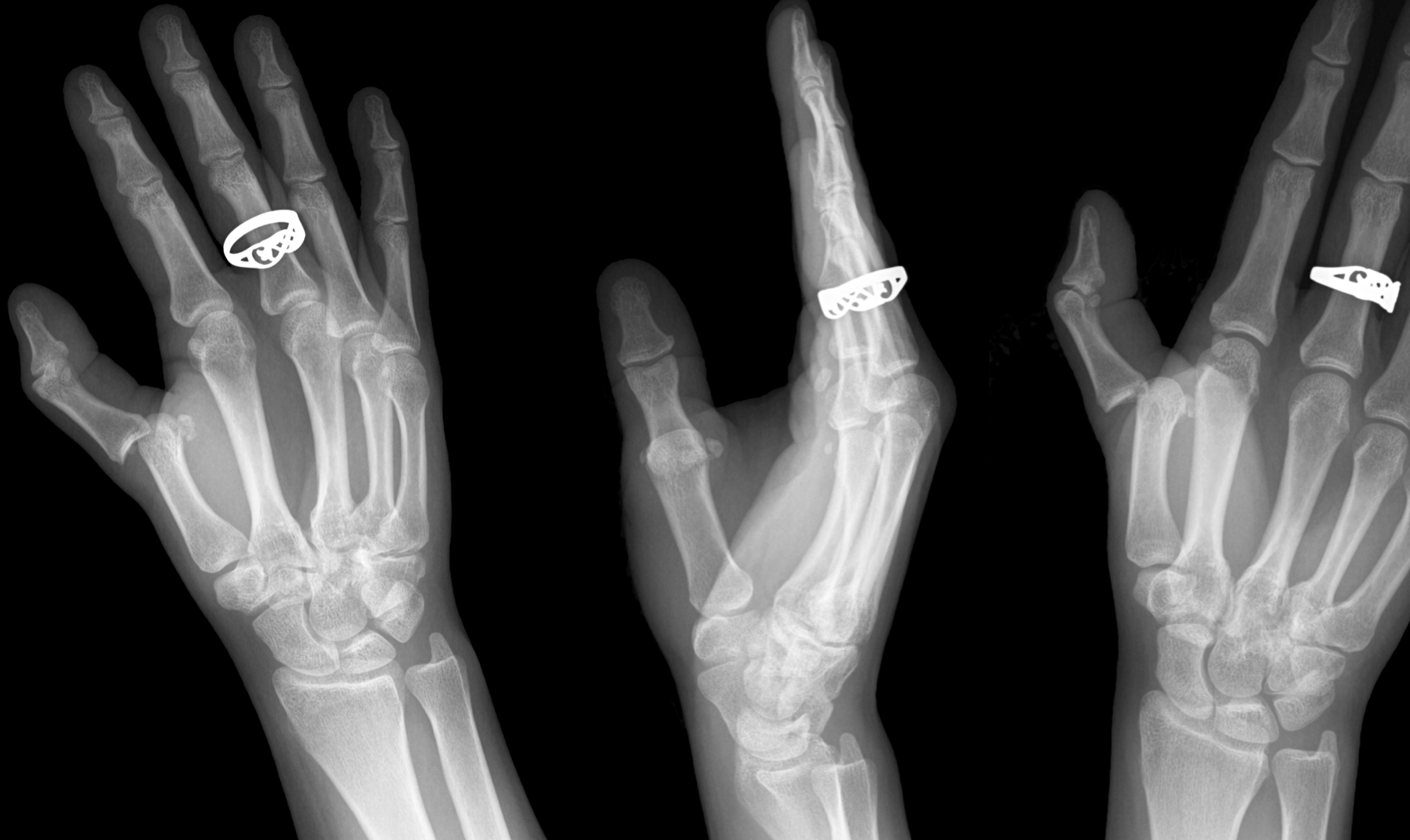
Dorsal Metacarpophalangeal (MCP) Dislocation
Follow me on Twitter (@criticalcarenow) or Google+ (+criticalcarenow)
Category: Visual Diagnosis
Posted: 9/29/2013 by Haney Mallemat, MD
(Updated: 9/30/2013)
Click here to contact Haney Mallemat, MD
65 year-old diabetic patient presents with abdominal pain. What's the abnormality on Xray?
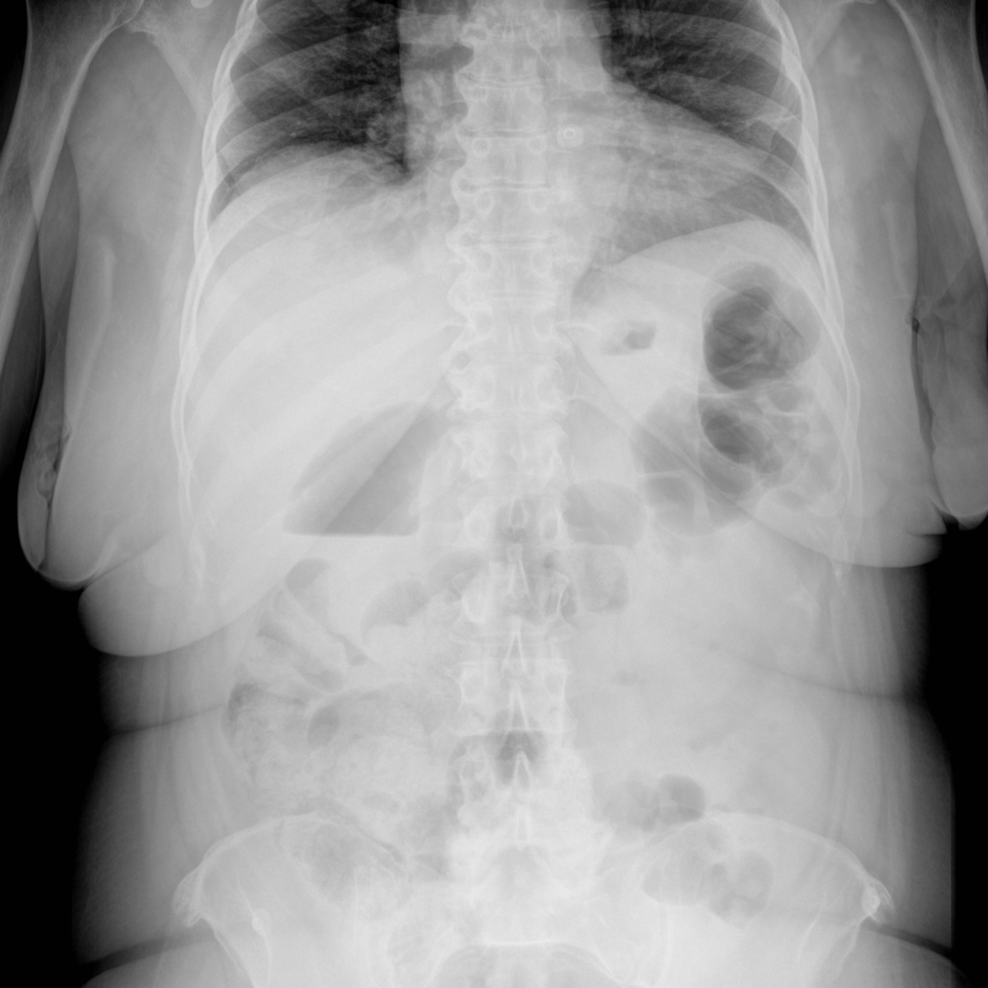
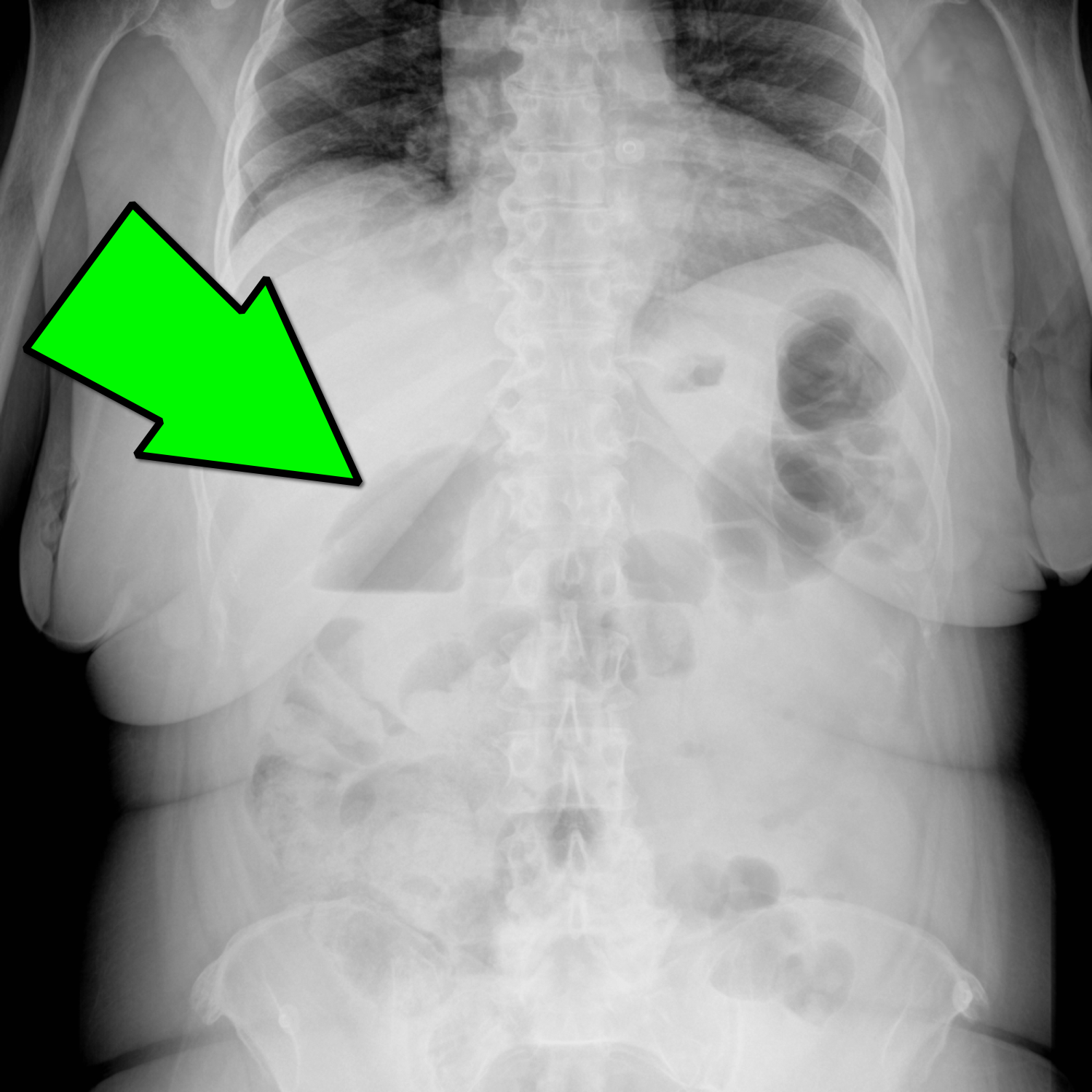
Emphysematous Cholecystitis
Emphysematous Cholecystitis
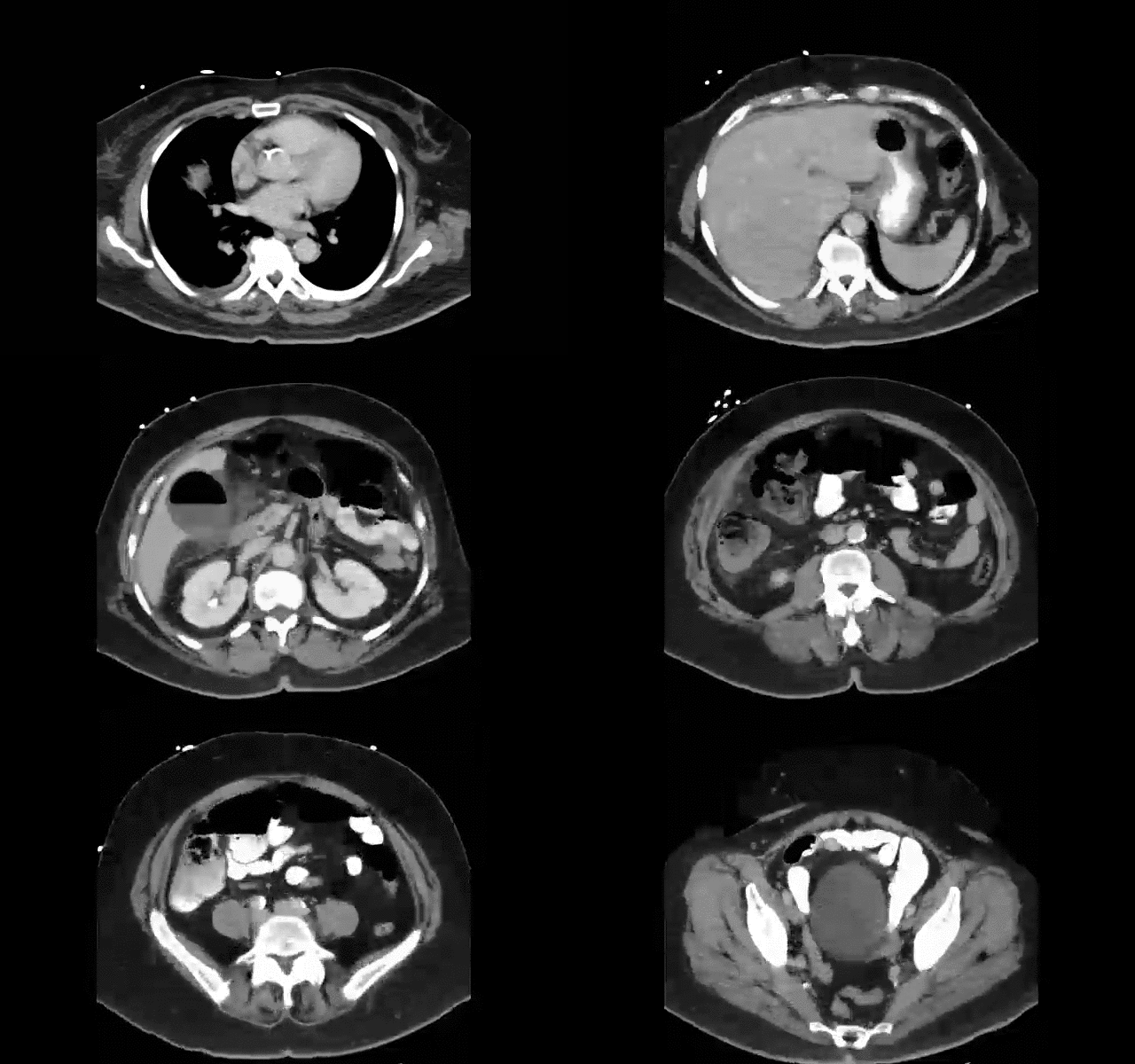
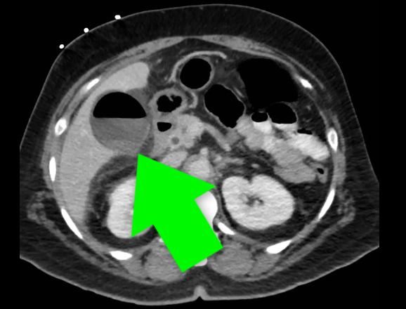
Carrascosa MF, et al. Emphysematous Cholecystitis. CMAJ.10 Jan 2012; 184(1): E81
Category: Visual Diagnosis
Posted: 9/23/2013 by Haney Mallemat, MD
Click here to contact Haney Mallemat, MD
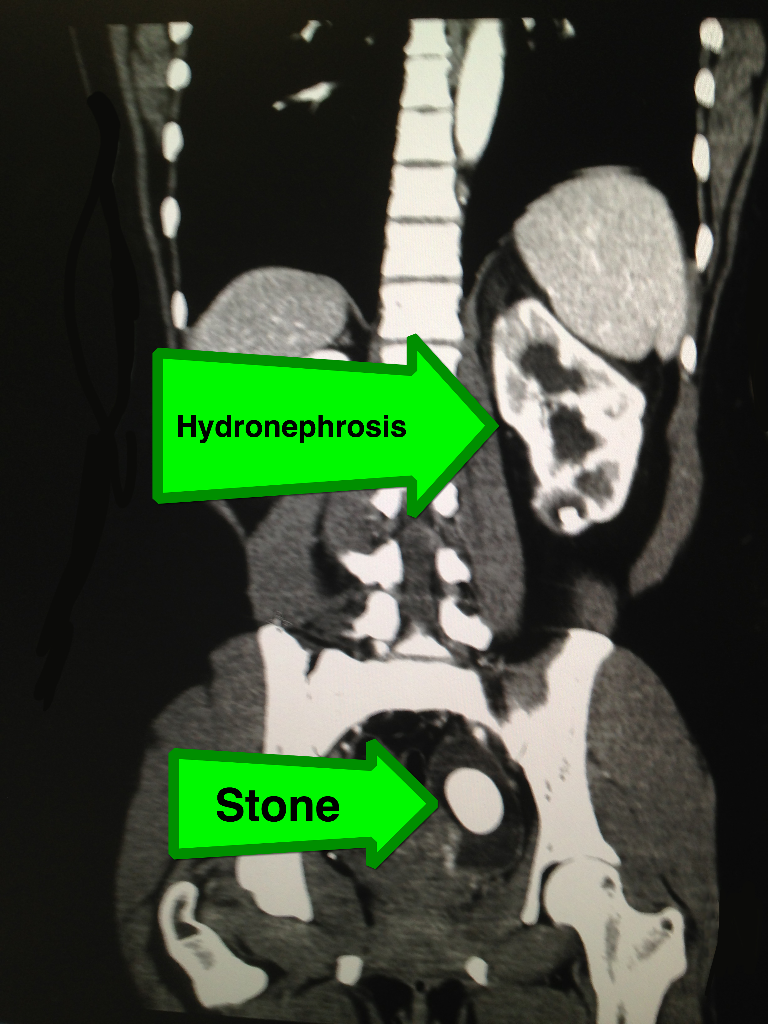
27 year-old female with no past medical history presents with sudden onset of left lower quadrant pain. What's the diagnosis?
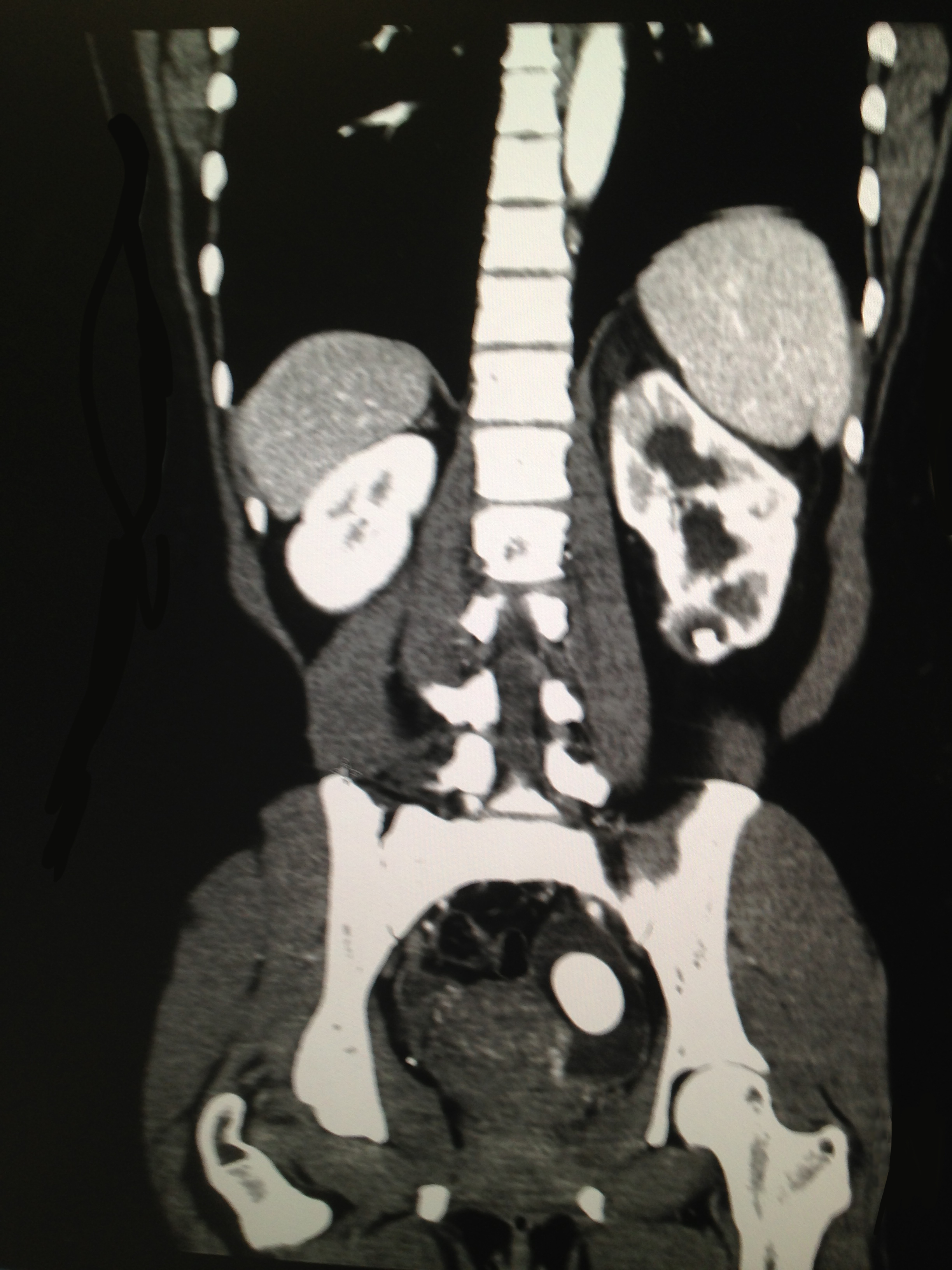
Large left-sided ureterolithiasis with hydronephrosis
Category: Visual Diagnosis
Posted: 9/16/2013 by Haney Mallemat, MD
Click here to contact Haney Mallemat, MD
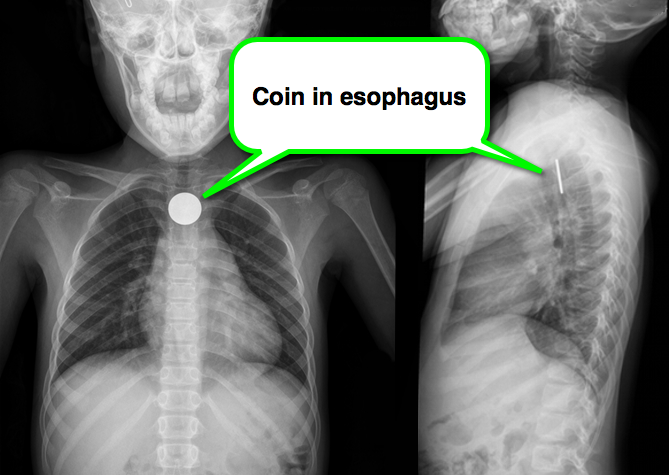
8 year-old girl presents with dysphagia and drooling, Xray is shown. What’s the diagnosis (and where is it located)?
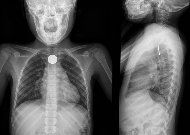
A coin located in the esophagus at the level of the cricopharyneus muscle
Foreign body (FB) pearls
Kenton, Foreign Bodies in the Gastrointestinal Tract and Anorectal Emergencies, Emerg Med Clin N Am 29 (2011) 369–400
Category: Visual Diagnosis
Posted: 9/8/2013 by Haney Mallemat, MD
(Updated: 9/9/2013)
Click here to contact Haney Mallemat, MD
This week's case is challenging, but very interesting...
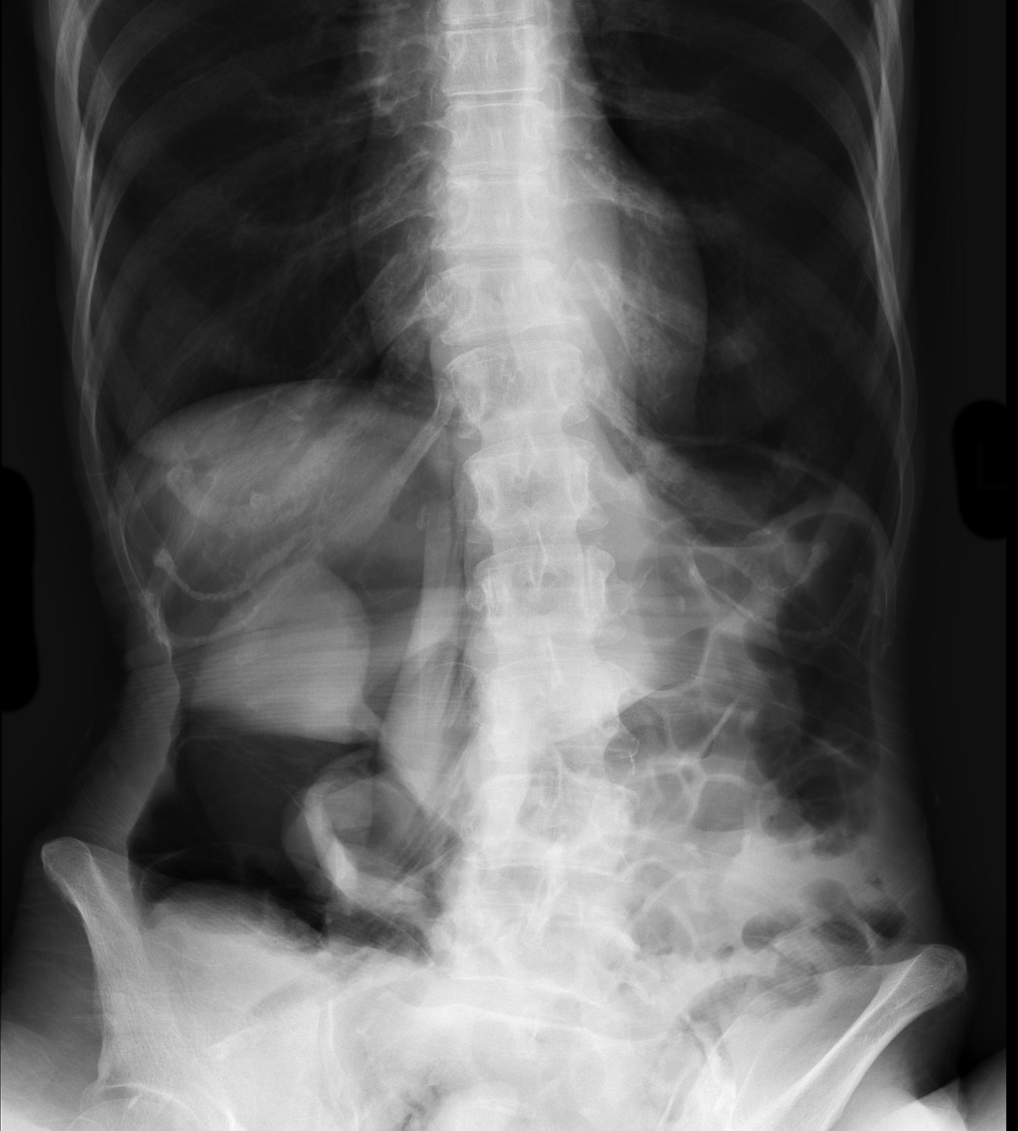
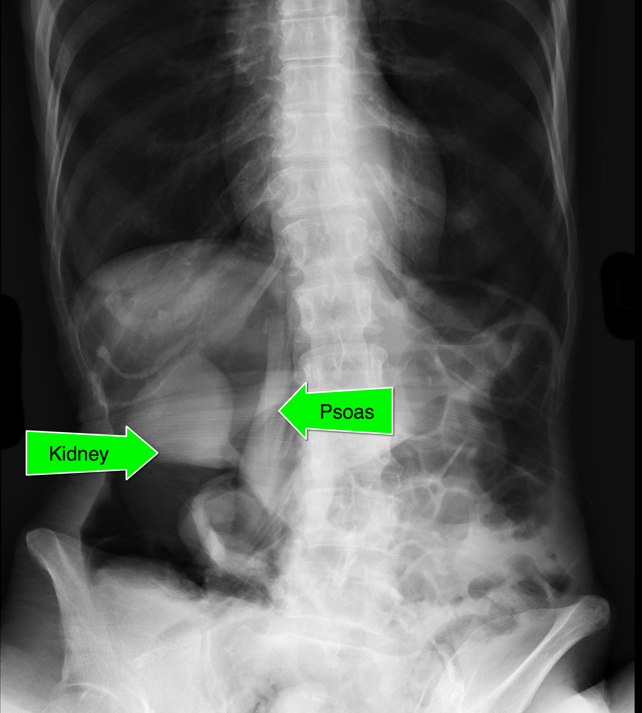
An elderly patient presents with a history of significant weight loss and chronic constipation; abdominal Xray is below. What's the diagnosis? (Hint: why is the right kidney and psoas muscle so well defined?)
Follow me on Twitter (@criticalcarenow) or Google+ (+criticalcarenow)
Category: Visual Diagnosis
Posted: 9/2/2013 by Haney Mallemat, MD
Click here to contact Haney Mallemat, MD
Elderly male presents with headache, confusion, and trouble with gait. What's in your differential diagnosis?
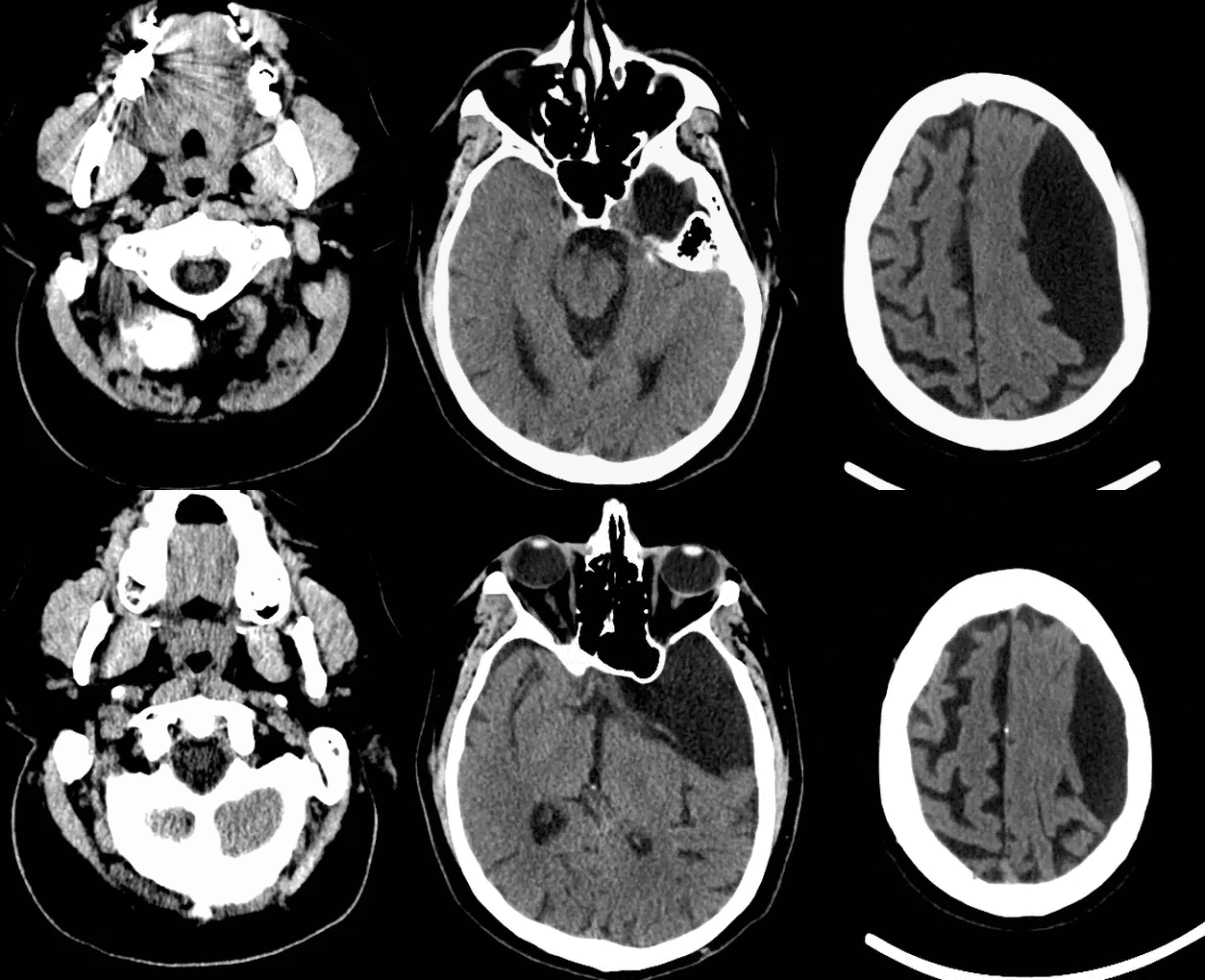
Based on the CT scan shown, the differential here includes epidermoid and arachnoid cyst
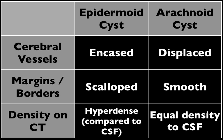
Arachnoid cysts (AC) occur within the cerebrospinal axis and do not communicate with the ventricular system. Most occur in the middle cranial fossa and are typically benign; continuing cerebrospinal fluid
The majority of AC occurs from abnormalities in development, but a small portion occurs secondary to post-surgical adhesions or in association with cancer.
MRI is the test of choice to help define the extent of the cyst as well as determine alternative diagnoses.
Treatment is variable with some experts stating that only symptomatic ACs should be treated with others recommending removal to avoid future complications.
The patient in the stem presented with symptoms secondary to complications from the AC.
Follow me on Twitter (@criticalcarenow) or Google+ (+criticalcarenow)
