UMEM Educational Pearls

Question

50 year-old male intubated for respiratory distress. Ultrasound is used post-intubation to confirm tube placement and the following images are obtained. What's the diagnosis?

Answer

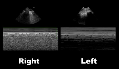
Right main-stem intubation as demonstrated by presence of lung-pulse on the left side

Lung-Pulse

  • Normal lung sliding creates an artifact at the pleural line, as the visceral and parietal pleura slide against one another; this is seen in the M-mode image below for the right lung
  • When a pneumothorax occurs the two pleural become separated and there is loss of this artifact; this is called the stratosphere sign (not shown here)
  • However, when the two pleura are in contact but there is no movement of the pleura (e.g. main-stem intubation on the non-ventilated side) lung-pulse is observed (seen below with arrows)
  • Although there is no ventilation of the affected lung, the lung pulse occurs because there is still cardiac activity that "pushes" the lung into the chest wall from underneath. This creates small periodic movements of the visceral and parietal pleura, creating small amounts of artifact
  • Lung pulse can differentiate pneumothorax from other causes of lung hypoventilation:
    • Mucus plug in main-stem bronchus
    • Foreign body in main-stem bronchus
    • Main-stem intubation

References

Follow me on Twitter (@criticalcarenow) or Google+ (+criticalcarenow)