UMEM Educational Pearls

Category: International EM

Title: What's the diagnosis?

Keywords: multiple myeloma, x-ray, global, neoplasm (PubMed Search)

Posted: 3/26/2014 by Andrea Tenner, MD
Click here to contact Andrea Tenner, MD

Question

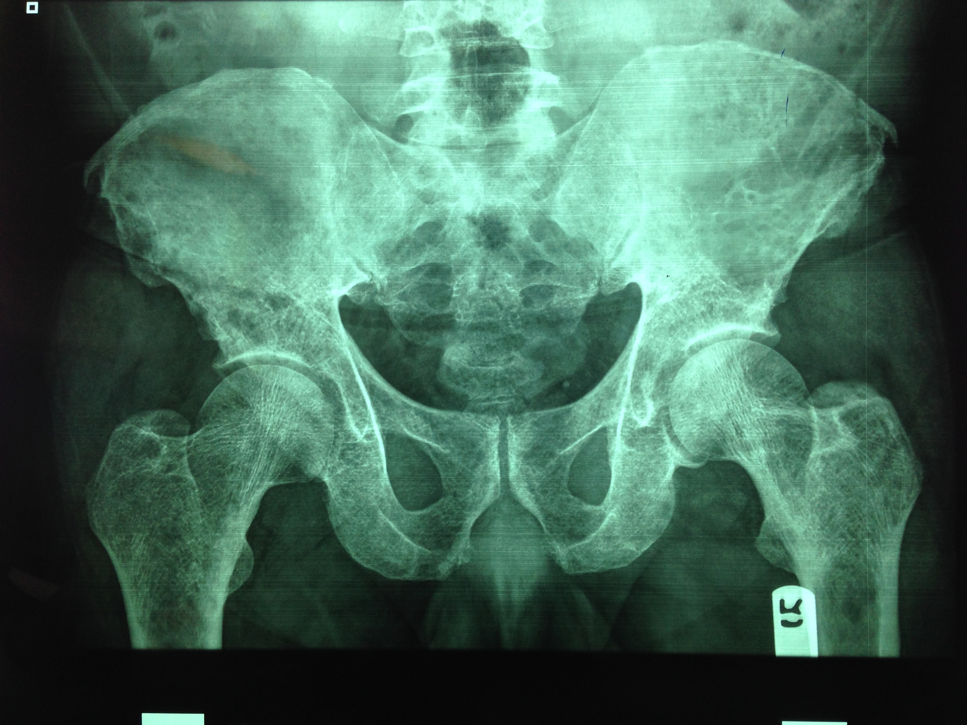
You are evaluating a 40 year old trauma victim and see this on pelvic xray. What are you worried about?

Answer

The patient likely has multiple myeloma. 
 
Multiple myeloma is a malignancy of plasma cells affecting the skeletal system. It has an osteoblastic effect on bones causing punched out lytic lesions in the skull, spine and pelvis. Skeletal survey is the initial diagnostic imaging of choice. It usually includes at least 3 of the following: a lateral radiograph of the skull, AP and lateral views of the spine, and AP views of the humeri, ribs, pelvis, and femora. It is also used as a staging tool to assess the extension of the disease.
 
Stage IA – Normal skeletal survey or single lesion
Stage IB - Five focal lesions or mild diffuse spine disease
Stage IIA/B - Five to 20 focal lesions or moderately diffuse spine disease
Stage IIIA/B - More than 20 focal lesions or severe diffuse spine disease
 
Treatment options are chemotherapy, radiation, steroids and stem cell transplant. Unfortunately, in patients with stage II these therapies are rarely curative.


University of Maryland Section for Global Emergency Health

Authors: Colleen Holley, MD and Van Pham, MD


References

Mulligan, Michael. Multiple Myeloma Imaging. available: http://emedicine.medscape.com/article/391742-overview#a19. accessed 24 March 2014.


Attachments

1403261757_IMG_0312.jpg (2,427 Kb)