UMEM Educational Pearls

Question

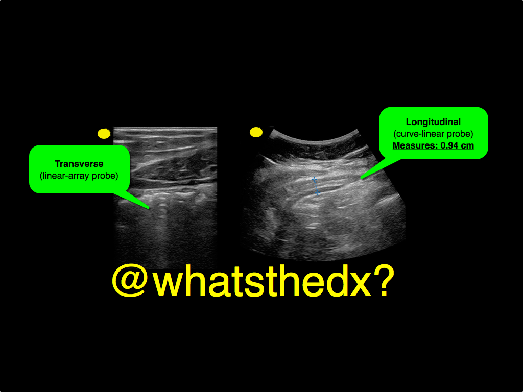
A male patient presents with right lower quadrant pain. The ultrasound is shown at the point of maximal tenderness. The diameter of the structure (image on right) is about 0.94cm. What is this structure and what's the diagnosis?

Answer

Appendicitis
  • Ultrasound can be used to diagnose appendicitis; a linear transducer is preferred in thinner / pediatric patients, while a curvilinear probe is may be used larger patients
  • Acute appendicitis causes irritation of the abdominal wall, so when looking for the appendix you need enough depth to visualize the peritoneal wall
  • To perform the scan, start at right upper quadrant in the transverse plane and slide down the abdomen towards the right iliac fossa until bowel gas has disappeared, typically the ileocecal junction. Alternatively, you can start scanning the point of maximal tenderness (i.e. the appendix)
  • Identifying a non-compressible and dilated appendix measuring greater than 6mm in transverse diameter suggesting the diagnosis
  • Color flow can be added to identify hyperemia around the appendix, which increases the specificity of the study.
  • More info on the scan can be found here and two podcasts from the incredible @ultrasoundpodcast guys part I and part II

References

Follow me on Twitter (@criticalcarenow) or Google+ (+criticalcarenow)