UMEM Educational Pearls

Question

A patient arrives in acute respiratory distress with left sided chest pain. Ultrasound of the left anterior chest is shown; what's the diagnosis and name one false positive?

Answer

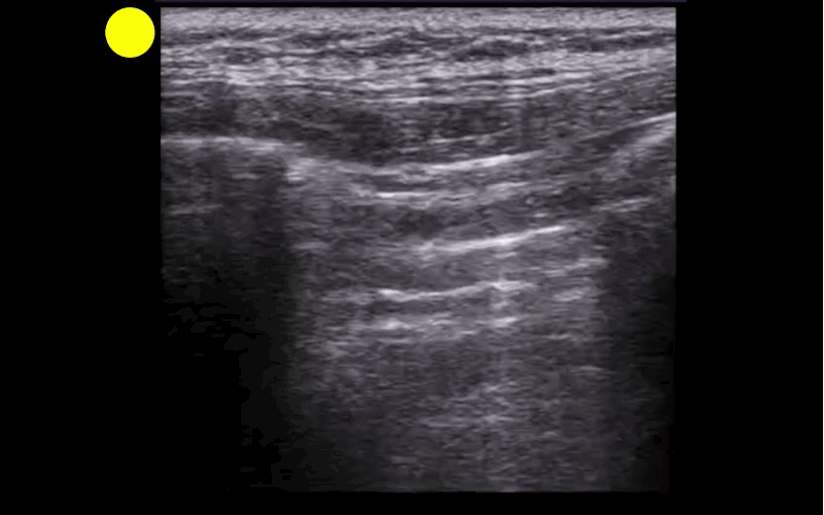
Lung point indicating pneumothorax (PTX)....see below for the false positives

What's the (Lung) Point

  • Separation of the visceral and parietal pleural secondary to a PTX leads to well recognized loss of lung sliding on ultrasound. 
  • Lung point (LP) on ultrasound is where the transition between pleural sliding and the loss of this sliding is demonstrated. Previous studies have reported that the LP has 100% specificity for a PTX.
  • Although this is a great sign to demonstrate PTX, be aware that there are several LP mimics:
    • lung-heart interface
    • lung-diaphragm interface (lower thorax laterally)
    • pleural effusion
    • blebs
  • Check out these great #FOAM posts by @ultrasoundjelly & @ultrasoundMD for more on the LP (and their mimics). Check them out here & here

References

Follow me on Twitter (@criticalcarenow)