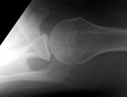
Pelllegrini-Stieda lesion
Ossified post-traumatic lesions at the MCL adjacent to the femoral attachment site of the medial femoral condyle.
Mechanism is likely from an avulsion injury that subsequently calcifies after the initial trauma.
Often an incidental finding on plain films.
If symptomatic, refer to ortho as an outpatient
If not symptomatic, no treatment is indicated
http://images.radiopaedia.org/images/30076/b62e61e83241e30f2da693901edcdc_gallery.jpg
http://www.imageinterpretation.co.uk/images/knee/PELLEGRINI%20STIEDA2.jpg
No single feature of the history of physical examination reliably rules out ostemyelitis
Aids in making the diagnosis include:
An ulcer area larger than 2 cm2 (LR 7.2),
A positive probe to bone test (LR 6.4),
An ESR greater than 70 mm/h (LR 11)
Treating knee osteoarthritis - from the American College of Rheumatology
Exercise whether it be aquatic, aerobic (land -based) or resistance can decrease pain and improve functional capacity. Exercise should be performed 3 to 5 times a week. Effects are usually noted after 3 to 6 months.
Weight loss of 5% or greater body weight is associated with a small improvement in pain and physical function. The main benefit of weight loss has more to do to effects on co-morbid conditions.
Walking aids: A single crutch or cane should be held on the side contralateral to the affected knee and should be advanced with the affected limb when walking to reduce the load on the affected joint.
Cane sizing: The distance from the floor to the patient's greater trochanter (brings the elbow to 15º to 20º of flexion.
Posterior Shoulder Dislocations
(A posterior shoulder dislocation will show the humeral head displayed superiorly in the image away from the clavicle which is the inferior most bone)
Some things to look for on the AP view that will suggest a posterior shoulder dislocation:
Life in the Fast Lane as a great discussion of posterior shoulder dislocations at http://lifeinthefastlane.com/posterior-shoulder-dislocation/
Best way to make the diagnosis --- suspect it and get an axillary view.
Unexplained respiratory symptoms during exercise are often incorrectly considered secondary to exercise induced asthma/bronchospasm.
An important diagnosis on the differential should be exercise-induced laryngeal obstruction (EILO).
Of 91 athletes referred for asthma workup, 35% had EILO.
The presence of inspiratory symptoms did not differentiate athletes with and without EILO.
61% of athletes with EILO used regular asthma medication at referral.
Compartment Syndrome
Compartment syndrome is classically described as having the 6 Ps:
The diagnosis of compartment syndrome can be difficult but ultimately it comes down to measuring the pressures in the area of concern. Various recommendations of the allowed pressure can be found, but in general a fasciotomy is not needed if the compartment pressure is 30 mmHg less then the diastolic pressure (The Delta 30). So if the patients diastolic pressure is 70, a fasciotomy is not need if the compartment pressure is less then 40.
Finally, if you are suspecting compartment pressure do NOT elevate the limb. Leave it in a dependent position to help improve blood flow into the limb.
Cauda equina syndrome results from compression of multiple lumbar and sacral nerve roots
Causes: Central disc herniation, spinal epidural abscess, malignancy, trauma, hematoma.
Consider this entity in those with back pain and radiculopathy at multiple spinal levels
Urinary retention occurs in >90% of patients
Saddle anesthesia occurs in 75%
Decreased rectal sphincter tone occurs in 60 to 80%
A post void residual volume <100 mL makes this entity very unlikely
Lateral hip pain
Findings of weakness and/or pain while testing hip abduction may point to gluteus medius muscle dysfunction with associated with greater trochanteric pain syndrome.
The Trendelenburg test may help. The patient stands on the affected leg. A negative test result occurs when the pelvis rises on the opposite side. A positive test result occurs when the pelvis on the opposite side drops and indicates a weak or painful gluteus medius muscle.
http://www.youtube.com/watch?v=TY-G4ErruUA
Prior fracture represents the strongest predictor of stress fracture in both sexes
For girls: Low body mass index, (<19), late menarche (age 15 or older), previous participation in gymnastics and dance.
For boys: increased number of seasons.
Participation in basketball appears protective in boys.
This may represent a modifiable risk factor for stress fractures.
The thumb MCP joint is subject to arthritric changes.
Sx's of arthritis will frequently present with pain in a similar region to deQuervain's disease.
The basal joint grind test
Perform by stabilizing the triquetrum with your thumb and index finger and then dorsally subluxing the thumb metacarpal on the trapezium while providing compressive force with the opposite hand.
http://www.youtube.com/watch?v=oEJH7KFGx_Y
Charcot Joint - Neuropathic arthropathy
A Charcot Joint is a progressive degeneration of a weight bearing joint that is normally seen in patients that have decreased peripheral sensation and proprioception.
Conditions associated with Charcot Joints are:
• Alcohol neuropathy
• Cerebral palsy
• Diabetes mellitus
• Spinal Cord Injury
• Strokes
• Syphilis (tabes dorsalis)
The foot is most commonly affected and radiographs can also show bony destruction, bone resorption, and gross deformity. The onset of pain and deformity is typically insidious. Charcot joints are often associated with ulcerations, secondary osteomyelitis, and can lead to amputations.
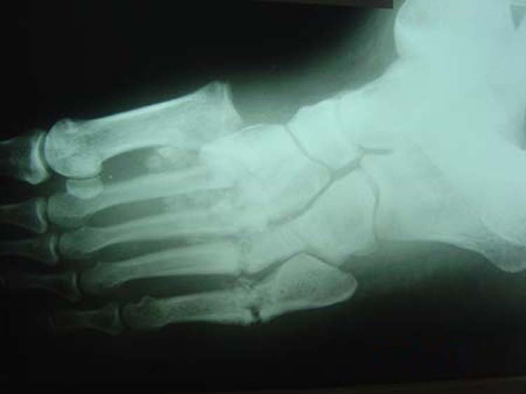

It is important to recognize the presence of a Charcot Joint so that the patient can be referred to Orthopaedics and treated (often with cast immobilization) to prevent further destruction of the joint.
The flexor tendons of the finger may become thickened and narrowed from chronic inflammation and irritation.
- Causes limitation in range of motion and snapping or locking during flexion
- Can involve any digit but usually the ring and the long finger
CC: pain, "catching" May awake to finger being "locked" with spontaneous resolution during the day
Stenosis occurs at the MCP level
PE: Distal flexor crease tender to palpation and may have a painful nodule
Full finger flexion is sometimes not possible
Tx: NSAIDs and steroid injection in tendon sheath. If this fails - surgical release.
Dupuytren disease is a nodular thickening and resultant contraction of the palmer fascia.
Increased in those of Northern European dissent.
One or more painful nodules located near the distal palmer crease.
Over time may result in flexion at the MCP joint.
Most commonly affects the ring finger.
Sensation is normal.
Over time affects ADLs
Tx: night splints and surgery
Tests for distal ulnar nerve entrapment
Ask patient to hold a piece of paper between the thumb and the index finger
Normally this is a fairly simple task.
With an unlar nerve palsy, the patient will substitute with the FPL (flexor pollicis longus - median nerve innervation). This causes flexion of the thumb in order to maintain the grip since the adductor pollicis cannot be used. This causes thumb flexion rather than extension.
http://www.mims.com/resources/drugs/common/CP0042.gif
http://www.youtube.com/watch?v=yJTIhm1VfSI
Sternal fractures
Tennis Elbow
The tendon usually involved in tennis elbow is called the Extensor Carpi Radialis Brevis (ECRB).
The ECRB muscle helps stabilize the wrist when the elbow is straight.
Ask the patient to straighten the arm at the elbow and then perform resisted long finger extension. This will stress the ECRB and reproduce the pain. One can also ask the patient to lift the top of a chair in the air with the elbow extended.
Trapezium Fractures
Suspect the Diagnosis when you note
If you are suspected the diagnosis oblique radiographs or a CT scan of the wrist will note the fracture the best.
Treatment consists of placing the patient in a thumb spica splint.
The adolescent brain has not yet reached full maturation and is in a period of rapid development from ages 14 - 16.
Adolescents have been found to be more sensitive to the effects of concussion than adults
Concussed adolescents have deficits in attention and executive function lasting up to 2 months post injury.
Be aware that the adolescent brain will require extended recuperation time following injury
In the future, discharge instructions might need to say more than "don't get hit in the head till your headache goes away." Because of deficits in attention and executive function, physicians should consider recommendations about adolescents and jobs, school work and driving an automobile.
Adhesive capsulitis aka frozen shoulder
idiopathic loss of BOTH active and passive motion (this is a significant reduction of at least 50%)
Motion is stiff and painful especially at the extremes
Occurs due to thickening and contracture of the shoulder capsule
Affects patients between the ages of 40 and 60
Diabetes is the most common risk factor
Imaging is normal and only helpful to rule out other entities such as osteophytes, loose bodies etc.
Treatment includes NSAIDs, moist heat and physical therapy.
Patients should expect a recovery period of 1-2 years!