Category: Orthopedics
Keywords: head injury, concussion, return to play, cognitive rest (PubMed Search)
Posted: 2/23/2013 by Brian Corwell, MD
Click here to contact Brian Corwell, MD
Just before you upgraded your old computer, recall what happened when you had Excel, Word and PowerPoint all open at the same time. In the concussed state, the brain is essenatially functioning like your old computer... and the more tasks it must perform, the slower it will work and slower it will recover. Hence the concept of cognitive rest. Below is taken from the AMSSM position statement of concussion in sport.
Return to school
There are no standardized guidelines for returning the injured athlete to school. If the athlete develops increased symptoms with cognitive stress, student athletes may require academic accommodations such as a reduced workload, extended test-taking time, days off or a shortened school day.
Some athletes have persistent neurocognitive deficits following a concussion, despite being symptom free. Consideration should be made to withhold an athlete from contact sports if they have not returned to their ‘academic baseline’ following their concussion (level of evidence C).
The CDC developed educational materials for educators and school administrators that are available at no cost and can be obtained via the CDC website. Additional resources for academic accommodations should be developed for both clinicians and educators (level of evidence C).
Adam Friedlander shared the practical application of this which I found amusing:
" I always recommend what Peds neuro called "a brain holiday" - my favorite part. All of our nurses look at me like I'm nuts, but it is now on our official concussion/CHI DC instructions. I always say to the kiddo: "You'll love this part. No homework, no reading." Then I turn to mom and dad and tell them they'll love the next part: "No TV, no video games."
Thank you for sharing Adam!!
American Medical Society for Sports Medicine position statement: concussion in sport
Category: Orthopedics
Keywords: head injury, concussion, return to play (PubMed Search)
Posted: 2/9/2013 by Brian Corwell, MD
(Updated: 2/1/2026)
Click here to contact Brian Corwell, MD
Estimated 3.8 million sport-related concussions per year (likely significantly higher due to underreporting)
Most patients recover within a 7-10 day period
** Children and teenagers require more time than college and professional athletes
This "accepted" time for recovery is not scientifically established and there is a large degree of variability based on multiple factors including age (as above), sex & history of prior concussions
Approximately 10% of athletes have persistent signs and symptoms beyond 2 weeks (which may represent a prolonged concussion or the development of post-concussion syndrome)
During this time the patient should have complete rest from all athletic activities, close follow-up with PCP and be educated re concussions.
If practical, "cognitive rest" should also be prescribed. This is one of the most frequently neglected aspects of post-concussion care and will be discussed in a future pearl.
Category: Orthopedics
Keywords: Hematoma Block, anesthesia, fracture reduction (PubMed Search)
Posted: 12/27/2012 by Brian Corwell, MD
(Updated: 2/1/2026)
Click here to contact Brian Corwell, MD
Hematoma Block
Provides good aesthesia for reduction of fractures. Onset in approximately 5 minutes
Benefits: No need for NPO, simple and easy to perform & can be done without additional personnel (unlike w/ procedural sedation)
Contraindications: Open fractures, dirty or infected overlying skin
1) Identify fracture site with x-ray and palpation
2) Clean skin w/ Betadine
3) Insert needle into the hematoma. * Confirm placement by aspirating blood *
4) Inject anesthetic (lidocaine 1 or 2%) into the fracture cavity and adjacent periosteum
http://www.youtube.com/watch?v=tjnsdjfwMmY
Category: Orthopedics
Keywords: head injury, concussion, return to play (PubMed Search)
Posted: 1/12/2013 by Brian Corwell, MD
(Updated: 2/1/2026)
Click here to contact Brian Corwell, MD
"When can my child get back out on the field doc?"
Return to play
▸ Concussion symptoms should be resolved before returning to exercise.
▸ A RTP progression involves a gradual, step-wise increase in physical
demands, sports-specific activities and the risk for contact.
▸ If symptoms occur with activity, the progression should be halted and
restarted at the preceding symptom-free step.
▸ RTP after concussion should occur only with medical clearance from a
licenced healthcare provider trained in the evaluation and management
of concussions.
Short-term risks of premature RTP
▸ The primary concern with early RTP is decreased reaction time leading
to an increased risk of a repeat concussion or other injury and
prolongation of symptoms.
Long-term effects
▸ There is an increasing concern that head impact exposure and
recurrent concussions contribute to long-term neurological sequelae.
▸ Some studies have suggested an association between prior concussions
and chronic cognitive dysfunction. Large-scale epidemiological studies are
needed to more clearly define risk factors and causation of any long-term
neurological impairment.
American Medical Society for Sports Medicine
position statement: concussion in sport, 2013
Category: Orthopedics
Keywords: metacarpal, neck, fracture (PubMed Search)
Posted: 12/29/2012 by Michael Bond, MD
Click here to contact Michael Bond, MD
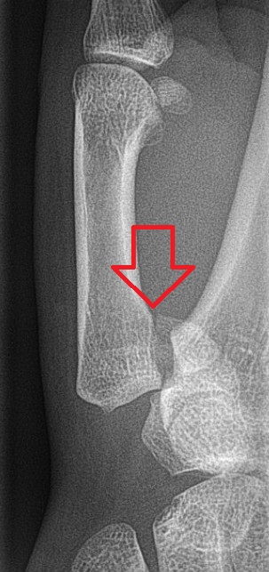
Metacarpal Neck Fractures (i.e.: Boxer’s Fracture if 5th Metacarpal)
Depending on the MCP joint involved a certain amount of angulation is permissible before it adversely affects normal function.
Wishing everybody a Happy and Healthy New Year.
Category: Orthopedics
Keywords: Exercise, NSAIDs, bowel injury (PubMed Search)
Posted: 12/22/2012 by Brian Corwell, MD
Click here to contact Brian Corwell, MD
NSAIDs are commonly used by professional and recreational athletes to both reduce existing and/or prevent anticipated exercise induced musculoskeletal pain
NSAIDs have potential hazardous effects on the gastrointestinal (GI) mucosa during strenuous physical exercise
Potential effects include mucosal ulceration, bleeding, perforation. and short-term loss of gut barrier function in otherwise healthy individuals
Intense exercise by itself has previously been shown to induce small intestine injury
Human intestinal fatty acid binding protein (1-FABP) is a protein found in mature small bowel enterocytes which diffuses into the circulation upon injury
Ibuprofen and endurance exercise (cycling) independently result in increased 1-FABP levels
When occurring together, ibuprofen ingestion with subsequent exercise causes significantly increased small bowel injury and intestinal permeability
Small bowel injury was found to be reversible in 2 hours
Taking empiric NSAIDs before endurance exercise may be an unhealthy practice and should be discouraged in the absence of a clear medical indication
Aggravation of exercise-induced intestinal injury by Ibuprofen in athletes. VAN Wijck K, Lenaerts K, VAN Bijnen AA, et al. Med Sci Sports Exerc. 2012 Dec;44(12):2257-62.
Category: Orthopedics
Keywords: pneumonia, rib fracture, blunt chest trauma (PubMed Search)
Posted: 12/7/2012 by Brian Corwell, MD
Click here to contact Brian Corwell, MD
Are discharged patients who suffer minor thoracic injury at risk of developing delayed pneumonia?
Prospective study of 1,057 patients age 16 and older with minor thoracic injury who were discharged from the ED.
32.8% had at least one rib fracture
8.2% had asthma
3.4% had COPD
Only 6 patients developed pneumonia!!
Sex, smoking, atelectasis on CXR, and alcohol intoxication were not significantly associated with delayed pneumonia.
However, for patients with preexistent pulmonary disease (asthma or COPD) AND rib fracture, the relative risk of delayed pneumonia was 8.6. Patients without either of these conditions are at extremely low risk of future development of pneumonia.
Patients with rib fractures do not develop delayed pneumonia: a prospective, multicenter cohort study of minor thoracic injury. Chauny JM, Emond M, Plourde M, Guimont C, Le Sage N, Vanier L, Bergeron E, Dufresne M, Allain-Boulé N, Fratu R. Ann Emerg Med. 2012 Dec;60(6):726-31.
Category: Orthopedics
Keywords: hematoma blocks, fracture analgesia (PubMed Search)
Posted: 11/24/2012 by Brian Corwell, MD
Click here to contact Brian Corwell, MD
Hematoma blocks for distal radius fractures
Hematoma blocks provide safe, effective analgesia without an increased risk of post procedural infections when compared with other regional blocks
Provide equal reduction quality AND pain control as procedural sedation with Propofol.
However, mean time to reduction (0.9 vs. 2.6 hours) and time to discharge post procedure (0.74 vs. 1.17 hours) were reduced with hematoma blocks.
Consider this option next time the department is busy or the patient is not an ideal procedural sedation candidate.
Emiley P, Schreier S and Pryor P. Hematoma Blocks for Reduction of Distal Radius Fractures. Emergency Physicians Monthly. September 2012. 16-17.
Category: Orthopedics
Keywords: tarsal tunnel syndrome (PubMed Search)
Posted: 11/17/2012 by Michael Bond, MD
(Updated: 2/1/2026)
Click here to contact Michael Bond, MD
Tarsal Tunnel Syndrome (TTS)
Prior pearls have addressed Carpal Tunnel Syndrome and Cubital Tunnel Syndrome, which affect the median and ulnar nerves, respectively. Tarsal tunnel syndrome, is a similar compression neuropathy of the tibial nerve as it transverses through the tarsal tunnel of the foot.
The tarsal tunnel is located behind the medial malleolus, and is where the posterior tibial artery, tibial nerve and several tendons transverse. Patients will present complaining of numbness of the foot radiating into Digits 1-4, pain, burning , and tingling of the base of the foot and heel. TTS has many causes and is more common in athletes.
Consider the diagnosis in patients with foot pain and numbness. If interested in more information about TTS please consider reading this eMedicine article, http://emedicine.medscape.com/article/1236852-overview
Category: Orthopedics
Keywords: calf tear, muscle tear (PubMed Search)
Posted: 11/10/2012 by Brian Corwell, MD
Click here to contact Brian Corwell, MD
Injury is often caused by sudden dorsiflexion on a plantar flexed foot w/ the knee in extension or similarly sudden knee extension with the ankle in a dorsiflexed position.
Injury has a predilection for the poorly conditioned middle-aged athlete, with "thick calves" who are engaged in strenuous activity
Strains are treated with ice, analgesics, and compression (decreases hematoma size and facilitates healing)
Also, consider casting/splinting as dictated by injury severity, such as with a night splint or a CAM boot.
Severe strains and ruptures can be splinted in plantar flexion for 3 weeks.
Gallo et al. Common Leg injuries of long-distance runners:Anatomic and Biomechanical approach. Sports Health. 2012
Category: Orthopedics
Keywords: fracture reduction, distal radius (PubMed Search)
Posted: 10/27/2012 by Brian Corwell, MD
Click here to contact Brian Corwell, MD
Distal radius fractures are common in children
Traditional management includes closed reduction +/- procedural sedation
The downside of this approach includes: patient risks, cost, physician time, ED bed time and tying up resources.
Kids have excellent bone remodeling potential...displaced and angulated fractures heal well without reduction
Crawford et al - 51 children aged 3 to 10 (avg 6.9 yrs) w/closed distal radius fractures.
Exclusions: open or growth plate fractures, metabolic bone disease or neurovascular injury.
No sedation, analgesia or fracture reduction was performed
Treatment: simple casting and gentle molding to correct angulation... i.e. fractures were left in a shortened, overriding position
Outcome: All patients had clinical and radiographic union and full range of motion of the wrist at one year w/ good patient (parent) satisfaction. This was associated w/ significant cost savings.
Consider this approach in consultation with orthopedist
Remember exclusions: open fractures, fracture dislocations, growth plate injuries and neurovascular injury.
Children w/ excessive angulation or rotational deformity should have standard care (closed reduction w/ sedation)
Multiple guidelines exist for "excessive angulation" but as a general rule
Age < 5 Up to 35 degrees
Age 5- 10 Up to 25 degrees
Age >10 Up to 20 degrees
Closed treatment of overriding distal radial fractures without reduction in children. Crawford et al.
J Bone Joint Surg Am. 2012 Feb 1;94(3):246-52.
Category: Orthopedics
Keywords: Marathon, cardiac arrest, cardiac death (PubMed Search)
Posted: 10/13/2012 by Brian Corwell, MD
(Updated: 2/1/2026)
Click here to contact Brian Corwell, MD
Congratulations to today's Baltimore marathoners and the medical race staff
In honor of them:
Marathons are becoming increasingly popular with participation rising from an estimated 143,000 US marathon finishers in 1980 to a record high of 507,000 during 2010.
Most victims of exercise-related sudden cardiac arrest have NO premonitory symptoms
Autopsy reports show that
1) 65 - 70% of all adult sudden cardiac deaths are attributable to coronary artery disease.
2) 10% due to other structural heart diseases (HOCM, congenital artery abnormalities)
3) 5 - 10% due to primary cardiac conduction disorders (prolonged QT, ion channel disorders)
4) Remainder are due to non cardiac etiologies
Overall risk of sudden cardiac arrest is approximately from 1 in 57,000 and the risk of sudden cardiac death is approximately 1 in 171,000. Mortality without intervention after sudden cardiac arrest is greater than 95%. The majority occur in middle to late aged males.
V fib/V tach are the most common arrhythmias leading to sudden cardiac arrest. Most events occur in the last 4 miles of the racecourse.
Survival decreases by 7 - 10% with each minute of delayed defibrillation. Defibrillation within 3 minutes can produce survival rates as high as 67 - 74%. After 8 minutes, there is a dramatic decrease in survival. Prompt CPR increases survival from 2.5% to greater than 8%.
Sudden cardiac arrest and death in united states marathons. Webner D, Duprey KM, Drezner JA, Cronholm P, Roberts WO. Med Sci Sports Exerc. 2012 Oct;44(10):1843-5.
Category: Orthopedics
Keywords: Fight, bite (PubMed Search)
Posted: 9/29/2012 by Michael Bond, MD
Click here to contact Michael Bond, MD
Fight Bites
Category: Orthopedics
Keywords: Shoulder, biceps, cartilage tear (PubMed Search)
Posted: 9/22/2012 by Brian Corwell, MD
(Updated: 11/19/2013)
Click here to contact Brian Corwell, MD
SLAP tear/lesion – Superior labral tear anterior to posterior
Glenoid labrum – A rim of fibrocartilaginous tissue surrounding the glenoid rim, deepening the “socket” joint and is integral to shoulder stability
http://www.orthospecmd.com/images/shoulder_labral_tear_anat_02.jpg
Injury is most commonly seen in overhead throwing athletes
Or from a fall on the outstretched hand, a direct shoulder blow or a sudden pull to the shoulder
Sx’s: A dull throbbing pain, a “catching” feeling w/ activity. Some describe clicking or locking of the shoulder. May also include nighttime symptoms. Pain is located to the anterior, superior portion of the shoulder.
Athletes may describe a significant decrease in throwing velocity
http://sitemaker.umich.edu/fm_musculoskeletal_shoulder/o_brien_s_test
Category: Orthopedics
Keywords: Apprehension test, patellar dislocation, (PubMed Search)
Posted: 9/8/2012 by Brian Corwell, MD
Click here to contact Brian Corwell, MD
Apprehension test for patellar dislocation
Test is used to access for the possibility of a patellar dislocation, prior to evaluation, now spontaneously reduced.
Similar to the shoulder apprehension test
Designed to place the patella in a position of imminent subluxation or dislocation
http://mulla.pri.ee/Kelley%27s%20Textbook%20of%20Rheumatology,%208th%20ed./HTML/f4-u1.0-B978-1-4160-3285-4..10042-7..gr16.jpg
http://www.youtube.com/watch?v=9AJxcbd9g8A
Place the knee in 20 - 30 degrees of flexion with the quadripces relaxed. Grasp the patella and attempt to place lateral directed stress.
If the patella is about to dislocate, the patient will experience apprehension due to the familiar pattern of dislocation, report the laxity and resist further motion by contracting the quadriceps
Category: Orthopedics
Keywords: shoulder dislocation, apprehension (PubMed Search)
Posted: 8/25/2012 by Brian Corwell, MD
(Updated: 2/1/2026)
Click here to contact Brian Corwell, MD
Apprehension test for shoulder dislocation
Tests for chronic shoulder dislocation
Similar to the patellar apprehension test
Designed to place the humeral head in a position of imminent subluxation or dislocation
http://www.maitrise-orthop.com/corpusmaitri/orthopaedic/112_kelly/kelly-fig11.jpg
ABduct and externally rotate arm to a position where the shoulder may dislocate
If the shoulder is about to dislocate, the patient will experience apprehension due to the familiar pattern of dislocation, report the laxity and resist further motion.
Category: Orthopedics
Keywords: lactate, synovial fluid, (PubMed Search)
Posted: 8/18/2012 by Michael Bond, MD
(Updated: 2/1/2026)
Click here to contact Michael Bond, MD
The Analysis of Synovial Fluid Analysis
When trying to diagnosis a septic joint, it is common to order the following labs on the synovial fluid:
Unfortunately, there is no value of glucose or protein that has enough sensitivity and specificity to make the tests diagnostically helpful. Gram stains are only postive in culture positive septic joints in approximately 50% of the cases. Cultures take too long to be helpful in the ED. The synovial WBC count can be helpful if very high, but a low value does not ensure that the patient does not have a septic joint.
The one test that has been shown to have a Positive Likelihood ratio of Infinity is a synovial lactate level >10. A synovial lactate should be sent on all synovial fluid as a level of 10 and greater makes the diagnosis of septic arthritis, regardless of the gram stain or synovial WBC level.
Carpenter CR, Schuur JD, Everett WW, Pines JM. Evidence-based diagnostics: adult septic arthritis. Acad Emerg Med. 2011 Aug;18(8):781-96.
Category: Orthopedics
Keywords: Humerus Fractures (PubMed Search)
Posted: 7/21/2012 by Michael Bond, MD
(Updated: 8/28/2014)
Click here to contact Michael Bond, MD
Humerus Fractures, Proximal
Category: Orthopedics
Keywords: Ulnar nerve, compression, neuropathy, wrist (PubMed Search)
Posted: 7/14/2012 by Brian Corwell, MD
(Updated: 2/1/2026)
Click here to contact Brian Corwell, MD
The median nerve is not the only compression neuropathy of the wrist
The ulnar nerve can become compressed at the level of the wrist as it 1) enters Guyon's canal or 2) or as the deep branch curves around the hook of the hamate
Compression can occur due to carpal bone fractures, local inflammation, ganglias, lipomas, anatomic abnormalities, etc
In sports medicine, the most common mechanism is injury is seen in cyclists (cyclist/handlebar palsy)
http://www.hughston.com/hha/b_15_3_2a.jpg
Also seen in those who participate in racquet sports, baseball, and golf
Symptoms can be isolated motor (claw hand = rare), sensory or both
http://en.academic.ru/pictures/enwiki/85/Ulnar_claw.jpg
Can be associated w/ median nerve compression
Tx: Activity modification such as wearing padded gloves, padding the object, or changing hand position on the handlebars
If above fails, surgical decompression is very effective.
Category: Orthopedics
Keywords: Bennett, Rolando, fracture (PubMed Search)
Posted: 6/30/2012 by Michael Bond, MD
(Updated: 2/1/2026)
Click here to contact Michael Bond, MD
First Metacarpal Fractures:
There are two types of fractures that commonly occur at the base of the 1st metacarpal. They are:
Bennett Fracture: This is an intraarticular fracture at the base of the 1st metacarpal that always involves some degree of subluxation or dislocation of the 1st carpometacarpal joint.

Image from Wikipedia Commons
Rolando Fracture: This is a communited intraarticular fracture at the base of the first metacarpal that typically has a T or Y shaped configuration with 3 fragments.

Image courtesy of WikiPedia Commons
