UMEM Educational Pearls - Visual Diagnosis

Category: Visual Diagnosis

Title: An alcoholic with fever and cough

Keywords: fever, cough, alcoholic (PubMed Search)

Posted: 5/7/2012 by Rob Rogers, MD (Emailed: 4/25/2024) (Updated: 4/25/2024)
Click here to contact Rob Rogers, MD

Question

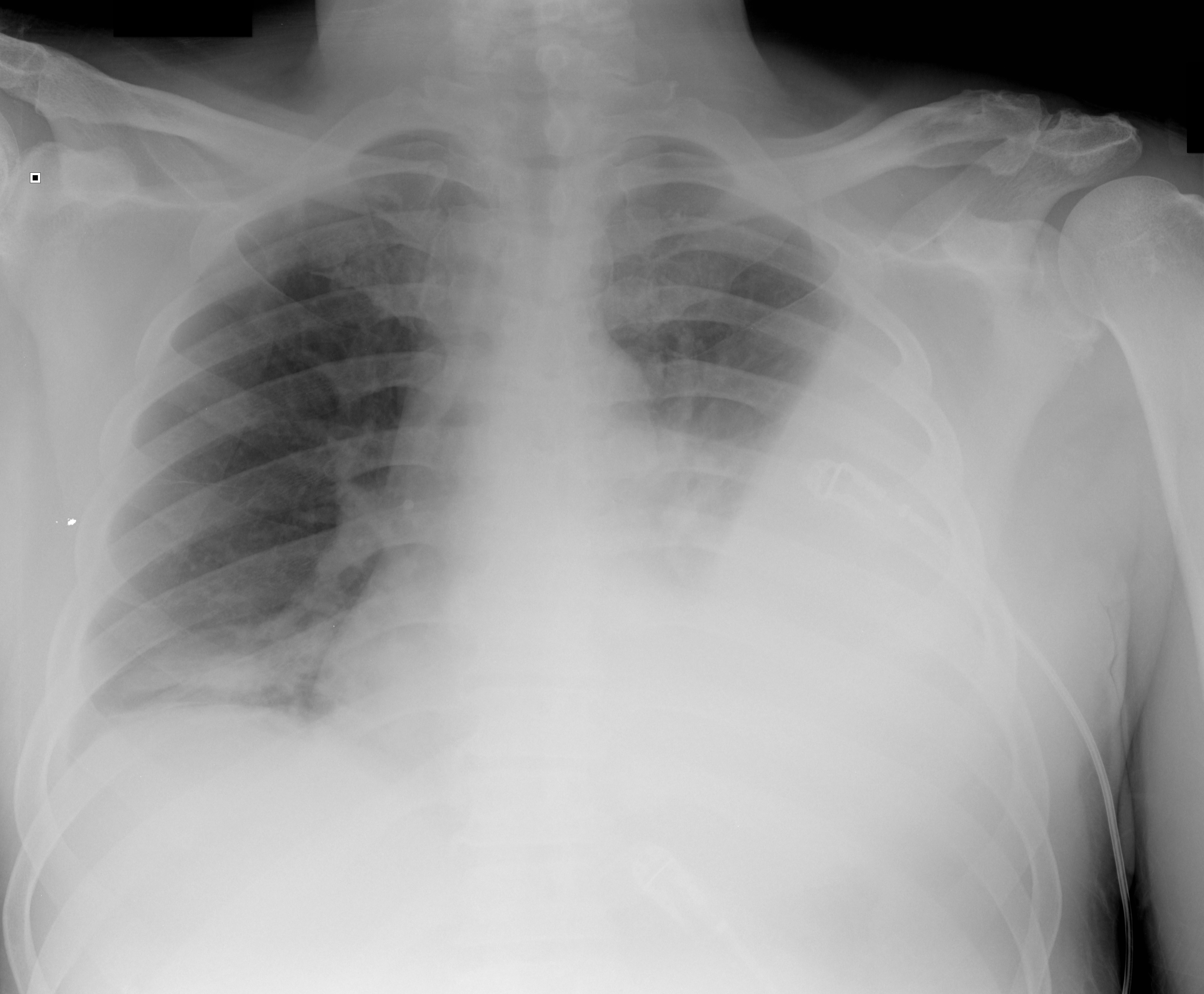
An alcoholic patient presents with a cough, fever, and very foul smelling breath (worse than usual)

What's the diagnosis? And what are the risk factors?


Show Answer

Show References


Question

50 year-old male with cough and dyspnea. What's the diagnosis?

Show Answer

Show References


Category: Visual Diagnosis

Title: What's the Diagnosis? Case by Dr. Ali Farzad

Posted: 4/7/2014 by Haney Mallemat, MD (Emailed: 4/25/2024) (Updated: 4/25/2024)
Click here to contact Haney Mallemat, MD

Question

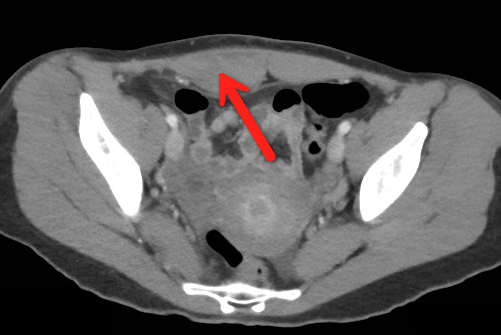
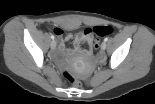
23 year-old female presents complaining of progressive right lower quadrant pain after doing "vigorous" pushups. CT abdomen/pelvis below. What’s the diagnosis? (Hint: it’s not appendicitis)


Show Answer


Category: Visual Diagnosis

Title: Visual Dx (Courtesy of Maite Huis in 't Veld)

Posted: 10/5/2018 by Michael Bond, MD (Emailed: 4/25/2024) (Updated: 4/25/2024)
Click here to contact Michael Bond, MD

Question

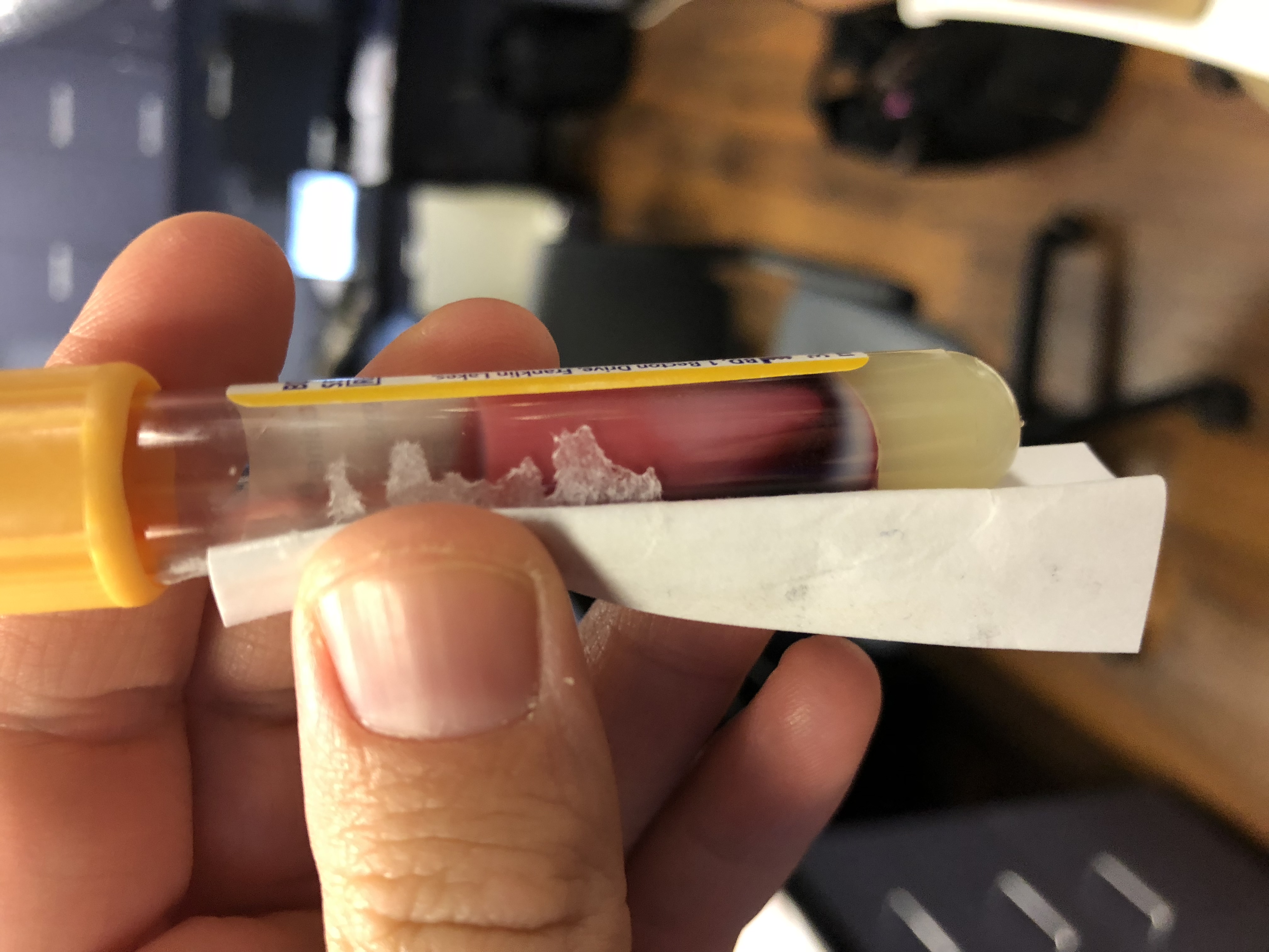
33 y/o M with PMH of ETOH induced pancreatitis presents with epigastic/RUQ pain & N/V after drinking last night, per patient his usual “pancreas pain”. The nurse shows you his blood tubes because they look “milky”. Lipase 1200, Ca 6.8.


What lab test would you add?


Show Answer


Category: Visual Diagnosis

Title: POCUS: Pulmonary Embolism (Submitted by Alexis Salerno)

Posted: 5/11/2022 by Michael Bond, MD (Emailed: 4/25/2024) (Updated: 4/25/2024)
Click here to contact Michael Bond, MD

Pulmonary Embolism 

  • In patients with high pretest probability and abnormal vital signs think about cardiac evaluation for pulmonary embolism. McConnell’s sign is most specific but can also be found in acute RCA infarct. TAPSE < 1.8 cm is also a good identifier of RV strain. Remember that patients with COPD or Pulm Htn may have RV dilation at baseline. You may also want to risk stratify patients with PE with labs as well as lower extremity dvt studies. 
  • Let’s give a shout out to Ashley Pickering who recently took some awesome echo images of a patient with a known saddle embolism. 


Show References