UMEM Educational Pearls

Question

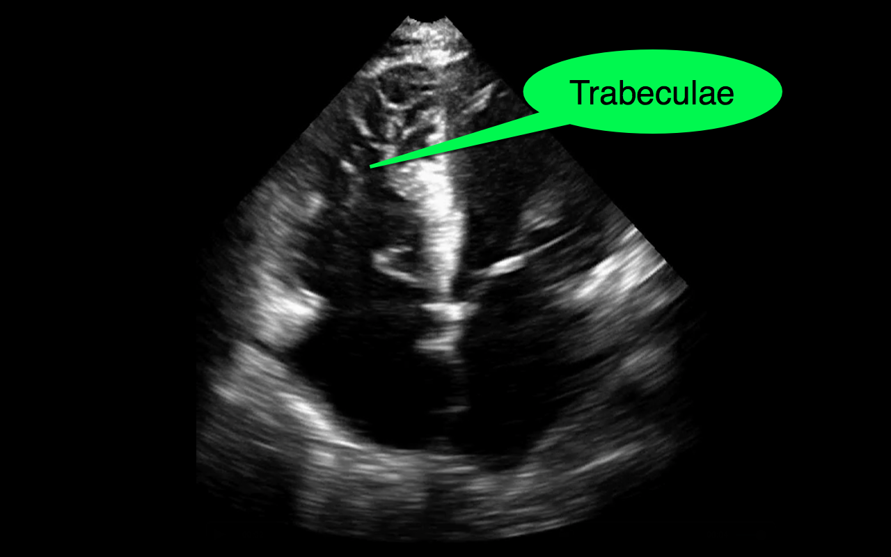
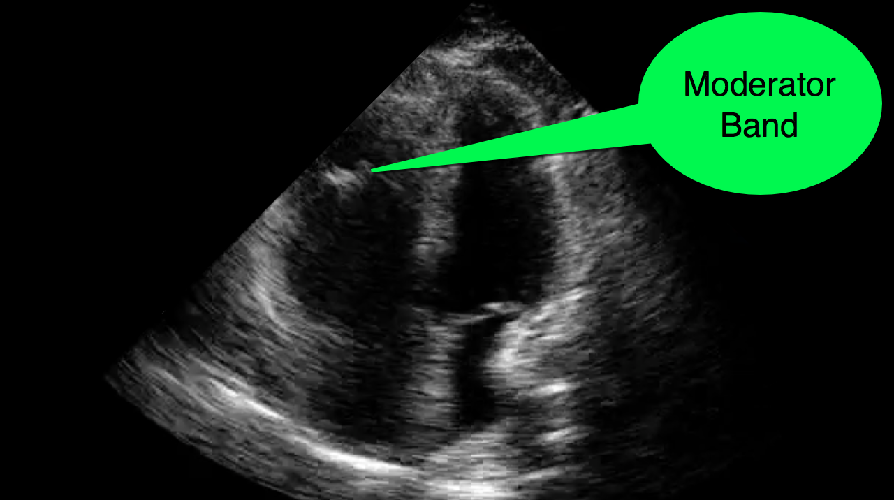
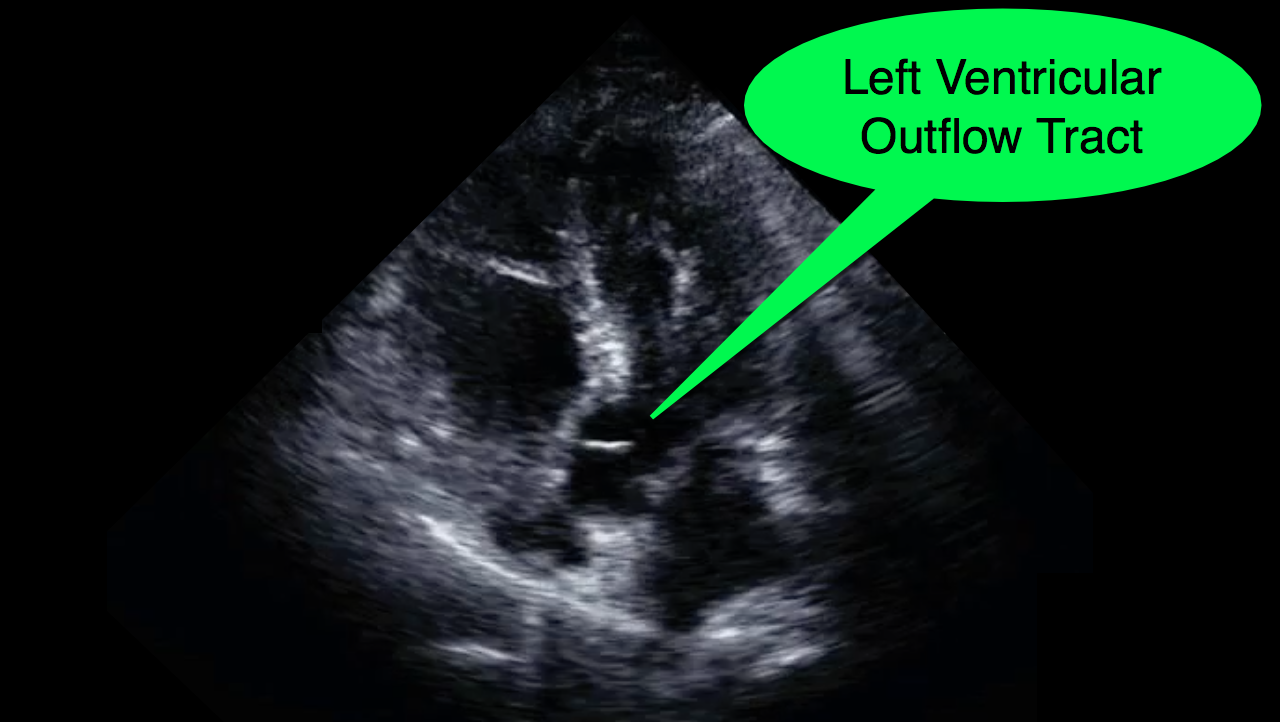
You decide to do a R.U.S.H. exam on your hypotensive patient and perform an apical four-chamber view.You see one of the two clips below; are there any tricks to figure out which is the left ventricle and which is the right ventricle?

Answer

Tricks to distinguish the RV from LV in apical four-chamber view

  • The right ventricle (RV) is normally smaller in size (2/3 the size) as compared to the left ventricle (LV).
  • When scanning critically ill patients you may encounter patients with right ventricles that are dilated secondary to acute or chronic illness. This enlargement may make the RV appear similar to a typical LV on an apical four-chamber view.
  • If you are new to ultrasound or just get confused by the probe marker, screen orientation, or just unsure, there are some tricks that you can use to quickly identify the LV and the RV on the apical four-chamber view.
    • The tricuspid valve is more "apically" displaced when compared to the mitral valve
    • The RV is heavily trabeculated as compared to the LV
    • The RV has the moderator band, while the LV lacks such a structure
    • The LV has the "fifth" chamber (i.e., the left ventricular outflow tract); you can identify this by tilting the probe slightly anteriorly after finding the apical four-chamber view.

References

Follow me on Twitter (@criticalcarenow) or Google+ (+criticalcarenow)