What are the signs of Cardiac Tamponade on ultrasound?
Think of them as a pyramid with clinical importance decreasing as you rise to the top of the pyramid.
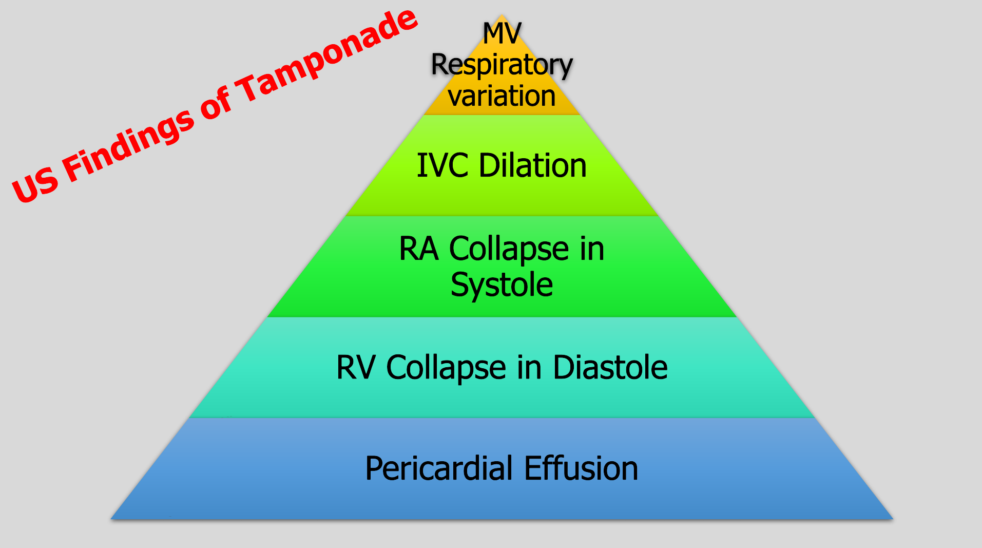
To have tamponade you need a pericardial effusion.
The most specific sign of tamponade is RV collapse in diastole.
The earliest and most sensitive sign is RA collapse over 1/3 of the cardiac cycle from late diastole into systole, which is why we say RA collapse during systole.
IVC dilation also occurs but is not sensitive.
Placing the pulse wave Doppler over the mitral valve and evaluating the change with respirations is an advanced technique. It’s positive if you have 25% change.
Don’t know if you are in systole or diastole? Connect your telemetry leads to the ultrasound machine. Don't have leads? Then you can also cine scroll on a subxiphoid view or parasternal view to look at when the valves are open and closed, then compare to the cardiac wall positioning.
Alerhand S, Adrian RJ, Long B, Avila J. Pericardial tamponade: A comprehensive emergency medicine and echocardiography review. Am J Emerg Med. 2022 Aug;58:159-174. doi: 10.1016/j.ajem.2022.05.001. Epub 2022 May 6. PMID: 35696801.