UMEM Educational Pearls - By Rob Rogers

Category: Vascular

Title: Follow-up for the Hypertensive Patient

Keywords: Hypertensive (PubMed Search)

Posted: 3/10/2009 by Rob Rogers, MD (Updated: 4/24/2024)
Click here to contact Rob Rogers, MD

Follow-up for the Hypertensive Patient

We see hypertensive patients every day, every shift. And, we discharge many of them. So, when do you get them follow-up?

The JNC-7 recommends that patients with BPs > 180/110 mm Hg have follow-up within 7 days. Like most of the HTN recommendations in the primary care setting, this recommendation is based on a "smart person" concensus....and no data.

This is a tremendous issue for us in the ED, because we don't want to see a bad outcome in our discharged hypertensive patients.

Some pearls regarding discharging the very hypertensive (but asymtomatic) patient:

  • Since there isn't any realy data on follow-up, it would be wise to use caution and have very high BPs checked the next day and to NOT wait a week.
  • Discharge instructions should note when/where (if you have to...use the ED as a recheck) the patient is to follow-up
  • ALWAYS warn patients about what can/will happen if they don't seek follow-up: MI, stroke, renal failure/need for dialysis, death, and disability and write this in the chart. The last thing you want to hear is that the patient went on to develop renal failure/stroke, etc. and that they claim they were not warned about what could happen.
  • When it is possible, contact the patient's doctor to discuss management


Category: Vascular

Title: Evaluation of End Organ Damage in Hypertensive Patients

Keywords: Hypertension, End-Organ Damage (PubMed Search)

Posted: 3/3/2009 by Rob Rogers, MD (Updated: 4/24/2024)
Click here to contact Rob Rogers, MD

Evaluation of End Organ Damage in Hypertensive Patients

No evidence to date supports the ED workup for end-organ damage in asymptomatic hypertensive patients.

End-Organ Damage Pearls:

  • Rarely, if ever, will an aimless search for lab abnormalities lead to any clinically meaningful change in patient management
  • An elevated creatinine does NOT define acute, end-organ damage. Most of the time it is due to the effects of chronic hypertension.
  • There is some evidence that a UA that has BOTH no protein and no red cells predicts a normal creatinine. The studies that have looked at this, however, are very small. Also, HTN in and of itself may cause some protein leak, even in the setting of normal renal function
  • A CXR and/or ECG is not needed in an asymptomatic patient.
  • Prompt followup is always necessary especially if no ED workup is started. All of this can be dome in the primary care doctor's office.


Category: Vascular

Title: Sudden onset thoracic back pain-think aortic dissection

Keywords: aortic dissection (PubMed Search)

Posted: 2/16/2009 by Rob Rogers, MD (Updated: 4/24/2024)
Click here to contact Rob Rogers, MD

BEWARE sudden onset thoracic back pain

Just reviewed a case last week of a person who presented with back pain (thoracic) as the sole manifestation of an aortic dissection. No chest pain, belly pain, etc. JUST severe, acute, thoracic back pain.

Keys to staying out of trouble:

  • Any sudden onset pain should be explained. Musculoskeletal pain doesn't normally present like this. Look for risk factors like HTN. If a person with HTN (even if not that high in the ED) presents with acute, severe, thoracic back pain the diagnosis of dissection should at the very least be considered.
  • The key to making the diagnosis begins with thinking about the diagnosis.
  • At the very least, include aortic dissection in EVERY patient you see with back pain, especially if sudden onset. I am not talking about the 95%+ people who don't really have anything wrong with them and who stumble into urgent care asking (begging) for Percocet.
  • Sudden onset back pain should also prompt consideration for a AAA
  • Just like all else in Emergency Medicine, always ask yourself if a "worst case scenario" could be present?, and the list for acute back pain is pretty short: dissection, AAA, fracture (by history), cancer, infection. Most of these, however, do not present acutely.

Show References


Category: Misc

Title: Pitfalls in ED Teaching

Keywords: ED Teaching (PubMed Search)

Posted: 2/10/2009 by Rob Rogers, MD (Updated: 4/24/2024)
Click here to contact Rob Rogers, MD

Pitfalls in ED Teaching

One of the best ways to improve as a teacher is to understand what mistakes expert educators have made in the past.

The following is a short list of pitfalls offered from some of the great teachers in our specialty:

  • Trying to teach for too long: "Teaching less is more"-that is to say, more will be remembered if the teaching session is brief.
  • Trying to teach too much: Trying to Stick to one main point, the "Educational Hit and Run," and move on
  • Failure to be enthusiastic when you teach: You must have some enthusiasm when you teach. Students/Residents won't learn as much or be as enthusiastic about learning without your enthusiasm!


Category: Vascular

Title: Pulmonary Embolism-Beware Two Important Atypical Presentations

Keywords: Pulmonary Embolism (PubMed Search)

Posted: 2/3/2009 by Rob Rogers, MD (Updated: 4/24/2024)
Click here to contact Rob Rogers, MD

Pulmonary Embolism-Beware Two Important Atypical Presentations

Seems like we have had several atypical PE presentations recently so I thought it timely to quickly highlight some of the well-reported presentations of pulmonary embolism. Remember, although we won't and can't diagnose every case, these types of presentations should at the very least prompt us to consider the diagnosis.

Atypical PE Presentations:

  • Syncope-occurs in as many as 15-20% of patients. Make sure PE is on the differential diagnosis of the syncopal patient, especially if there was any preceeding shortness of breath or chest pain.
  • Abdominal pain-we just had a case of this last week. A young female 6 weeks into a course of OCPS developed RUQ pain that radiated to the left shoulder. She had NO shortness of breath. However, the RUQ pain was pleuritic. Remember the movement of the diaphragm as it is responsible for abdominal pain presentations of both PE and pneumonia. A d-dimer was obtained and returned 3000. A CT scan was then ordered which showed a large right lower PE. What's the moral of the story? Well, it isn't to rule out PE in patients with belly pain. The lesson here is that upper abdominal pain may reflect disease in the chest (lower lobe pneumona and PE) and vice versa. To make matters worse an ultrasound of the RUQ was ordered 1st which showed gallstones!


Category: Misc

Title: Feedback as a Teaching Tool

Keywords: Feedback, Teaching (PubMed Search)

Posted: 1/26/2009 by Rob Rogers, MD (Updated: 4/24/2024)
Click here to contact Rob Rogers, MD

Feedback as a Teaching Tool

Why do we, in general, stink at giving feedback?

  • We were never taught how to do it
  • We fear we will hurt someone's feelings
  • It's painful to give feedback

Consider a few quick pearls that will increase your success at giving valuable feedback:

  • Realize that learners (students/residents) crave feedback....proven in multiple studies
  • Feedback IS a powerful teaching tool and isn't just a way of evaluating someone.
  • Avoid at all cost, the phrase,"good job." Be specific about what you mean
  • Praise in public, perfect in private
  • Avoid the "complain syndrome" and don't fall victim to it. This refers to the phenomenon in which we complain about a behavior or trait and NEVER actuall tell the person. We have all done it. Set yourself apart from others by giving the learner the needed feedback.
  • Learners won't improve without feedback. Just like the Nike commercial says,"Just do it!"


Category: Misc

Title: Teaching in the Emergency Department

Keywords: Teaching, Emergency Department (PubMed Search)

Posted: 1/20/2009 by Rob Rogers, MD (Updated: 4/24/2024)
Click here to contact Rob Rogers, MD

Teaching in the Emergency Department

Effective ways to teach in the ED:

  • Limit the amount of time you spend teaching (more teaching does not = more learning)....Take Home Point: teach a quick pearl about a case and move on. Dont belabor the point and keep teaching for 5-10 minutes. You will loose the learner.
  • Make teaching points applicable to the patient. Theoretical stuff is fine but no one cares about the Krebs cycle or ATP.
  • Teach "on the fly" (teach as good teaching moments come up on each case). "Board talks" are nice but are often times not practical in a busy ED.
  • Above all, be enthusiastic...without this all teaching will be ineffective

Show References


Thrombolytic Therapy for Pulmonary Embolism

Indications for administration of fibrinolytic therapy for acute PE:

  • Cardiac arrest presumed to be secondary to PE-tPA 50 mg bolus, may be repeated once.
  • Massive PE (hemodynamic instability)-arbitrarily defined by BP < 90 mm Hg systolic. Give 10 mg tPA bolus followed by 90 mg over 2 hours. Make sure heparin off during this time frame. tPA is the only FDA approved drug for this but some are starting to use Tenecteplase (single 0.5 mg/kg bolus).
  • Submassive PE (normal hemodynamics and evidence of RV strain). This tends to be the most controversial group, although many authorities are now advocating its use. Strongly suspect strain if the Troponin/BNP are elevated and get an ECHO if they are. Most studies that advocate for lytics in this group show significant improvement in PA pressures, RV wall dilatation, etc. What is currently missing is outcome data...i.e. how short of breath and disabled are people with submassive PE at 6, 9, and 12 months? Bottom line, enough evidence exists to support giving to stable patients with RV strain as long as they are carefully screened.
  • There is NO evidence that lytics are useful in stable patients without RV strain.
  • The administration of thrombolytic therapy for acute PE is within the scope of practice of emergency medicine.


Show References


Category: Vascular

Title: Neurologic Manifestations of Acute Aortic Dissection

Keywords: Acute, Aortic Dissection, Neurologic (PubMed Search)

Posted: 1/6/2009 by Rob Rogers, MD (Updated: 4/24/2024)
Click here to contact Rob Rogers, MD

Neurologic Manifestations of Acute Aortic Dissection

A myriad of neurologic presentations of acute aortic dissection have been reported in the literature. Although classic CVA symptoms may occur, nonspecific neurologic symptoms are much more common

These include:

  • Classic stroke-like/TIA symptoms
  • Encephalopathy (may look like a drug overdose)
  • Seizures (ask Mike Abraham about his abdominal pain/seizure case)

Take Home Point:

  • Consider the diagnosis of acute aortic dissection in patients with these findings who ALSO HAVE chest, back, or abdominal pain +/- risk factors for the disease (i.e. HTN, family history, Marfans, cocaine, etc.)

Show References


Category: Infectious Disease

Title: Infections That Cause Temperature-Pulse Dissociation

Keywords: Infections, Temperature (PubMed Search)

Posted: 12/29/2008 by Rob Rogers, MD (Updated: 4/24/2024)
Click here to contact Rob Rogers, MD

This pearl is dedicated to Dr. Michael Rolnick....


Infections That Cause Temperature-PulseDissociation

Certain infections may cause temperature-pulse dissociation (relative bradycardia in association with fever).

Remember that normally there will be an increase in pulse rate by 10 bpm for every 1 degree increase in temperature. So, if a patient has a temperature of 103 F, expect them to be tachycardic.

Any intracellular organism has the potential to cause a relative bradycardia (Faget's sign)

Infections that cause dissociation:

  • Salmonella typhi
  • C burnetii (agent of Q fever)
  • Chlamydia infections
  • Dengue fever


Category: Hematology/Oncology

Title: Typhlitis

Keywords: Neutropenic Entercolitis (PubMed Search)

Posted: 12/22/2008 by Rob Rogers, MD (Updated: 4/24/2024)
Click here to contact Rob Rogers, MD

A neutropenic cancer patient that presents with right lower quadrant abdominal pain, fever, and bloody diarrhea should raise suspicion for typhlitis (necrotizing colitis, cecal inflammation). This most commonly occurs in patients with hematologic malignancies who have been treated with cytotoxic agents. This condition is high risk and is associated with high morbidity and mortaiity.

Treatment:

  • Broad-spectrum antibiotics
  • CT scan of the abdomen and pelvis
  • Surgical consultation
  • Usually requires ICU admission

Show References


Category: Med-Legal

Title: Chest Pain Documentation

Keywords: Chest Pain (PubMed Search)

Posted: 12/15/2008 by Rob Rogers, MD (Updated: 4/24/2024)
Click here to contact Rob Rogers, MD

There is clearly no way you can document everything on a chest pain chart. However, there are some pretty important things that should be on the chart.

Some key things to consider documenting:

  • Why you did not work up someone's chest pain, i.e. what would you want your chart to look like if the patient went home to have an MI and an attorney looked at your chart? You don't think a ECG is warranted? Fine. Just document why. The chart tells all.
  • Documentation of risk factors for the three deadly causes of chest pain: ACS/MI, aortic dissection, and PE. Documenting these is proof you were thinking about a differential diagnosis.
  • Documenting key chest pain physical exam findings and pertinent negatives-Documenting "legs normal, no DVT" is proof you were thinking about PE the whole time, even if it isn't in your medical decision making section. Writing "no diastolic murmur" is proof you thought about aortic dissection. These kinds of documentation pearls will serve to make the chart defensible. Obviously, you should perform this part of the exam and not just write it on the chart.
  • Documentation of why you didn't go after ACS, aortic dissection, or PE. We will all make mistakes in our careers. And remember, we can't diagnose every MI, dissection, and PE. But, remember that you want your chart to show that you thought about these bad boys and WHY you didn't go after them. What is frequently missing on charts of missed MI, AD, and PE is exactly this!

Show References


Category: Vascular

Title: Hypertension and Epistaxis

Keywords: Hypertension, Epistaxis (PubMed Search)

Posted: 12/8/2008 by Rob Rogers, MD (Updated: 4/24/2024)
Click here to contact Rob Rogers, MD

Hypertension and Epistaxis

We commonly encounter patients with epistaxis who are found to be hypertensive. Some have taught over the years that hypertension causes nosebleeds and that some nose bleeds won't stop until the BP is lowered...

Some pearls about HTN/Epistaxis:

  • Most patients we see with hypertension are not experiencing epistaxis, casting serious doubt on a causal relationship
  • Studies show that the degree of blood pressure elevation does not correlate with risk of nose bleed
  • No studies have ever shown that acute BP reduction in the ED for a nose bleed is beneficial or reduces bleeding
  • Much of the debate is sparked by our ENT colleagues who swear that hypertension leads to nose bleeds and that bleeding will not stop until the BP is "treated." Much of this is based on experience with patients in the OR or IR suite. These blood pressures tend to be treated with IV antihypertensives by the ENT folks, and they feel pretty strongly about this relationship.


Category: Vascular

Title: Warfarin Induced Skin Necrosis

Keywords: Warfarin, Skin Necrosis (PubMed Search)

Posted: 12/1/2008 by Rob Rogers, MD (Updated: 4/24/2024)
Click here to contact Rob Rogers, MD

Warfarin-Induced Skin Necrosis (WISN)

Some pearls about a rare, but serious side effect of Warfarin...

  • WISN Occurs in 0.01-0.1% of patients taking Warfarin
  • More common in middle-age, perimenopausal women being treated for DVT/PE
  • Symptoms usually begin on days 3-6 of Warfarin treatment
  • Underlying pathophysiology is complex but involves thrombosis of superficial dermal capillaries
  • Postulated to be associated with deficiencies of protein C, protein S, and antithrombin III
  • Rash is most common on the breats, with thighs/buttocks being second most common site (see picture)
  • Diagnosis usually made clinically based on appearance of rash
  • Treatment is aimed at restoring Vitamin K dependent clotting factors by administering Vit K and FFP
  • For patients with the need for anticoagulation (DVT/PE, etc.) Heparin therapy is usually started


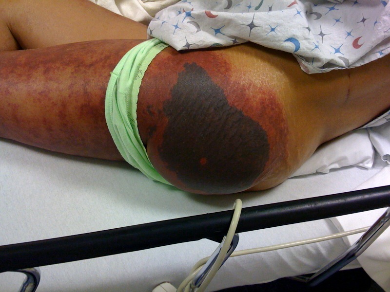
55 yo female presented to the ED on the day of hospital discharge for evaluation of this rash.

The rash began 4 days after starting Warfarin. Was being treated for a DVT.


Attachments

0812012217_photo.jpg (137 Kb)


What Hypertensive Patient Needs a Workup for End-Organ Damage?

Ah, the age old question...which hypertensive patients need an ED workup for end-organ damage? The "workup" for patients includes renal function, urinalysis, CXR, ECG, etc.

Some pearls regarding working patients up: 

  1. Asymptomatic patients in general do not need a workup. There is pretty good literature that shows you just won't find much (expecially anything that will change your treatment plan) if you go hunting in this group of patients.
  2. If you set asymptomatic patients aside, you won't find much good data on how much of a workup other patients need. Does a 45 yo patient with a BP of 160/110 and a mild HA need a serum creatinine? What if they have had some mild, atypical CP? The answer is...no one knows. Much of what we we do depends on what we were taught and our current mood. 
  3. Asymptomatic patients (truly asymptomatic) don't need chest xrays and ECGs as a rule of thumb...what you find won't help you make a decision. If you find LVH on the ECG, so what? 
  4. Obtaining a serum creatinine makes sense, especially of you are going to start a BP agent. 
  5. There is a pretty good study by Karas, et al. that showed that a urinalysis without protein or blood predicts a normal creatinine. Use caution, however, if you use this as a screen for renal disease, because many patients with HTN spill protein (despite a normal creatinine)


Category: Infectious Disease

Title: Healthcare Associated Pneumonia

Keywords: Pneumonia (PubMed Search)

Posted: 11/18/2008 by Rob Rogers, MD (Updated: 4/24/2024)
Click here to contact Rob Rogers, MD

Healthcare Associated Pneumonia (HCAP)....why is this important for the emergency physician?

Most of us are very familiar with the types of pneumonias commonly seen in clinical practice: community-acquired pneumonia (CAP), hospital-acquired pneumonia(HAP), and ventilator-associated pneumonia (VAP). But, some may not be that aware of a relatively newer type of pneumonia that has been well-defined, healthcare-associated pnemonia (HCAP). Experts in infectious disease and critical care now say that we (the ED) should be assessing ALL pneumonia patients for HCAP risk factors.

Why care, you ask?

  • Higher mortality than CAP
  • May look like CAP
  • Treated much differently than CAP

Risk factors: (most are common sense)

  • Nursing home or extended care facility resident
  • Recently admiited to a hospital for 2 or more days in the preceeding 90 days
  • Home wound care or attending a clinic for wound care
  • Dialysis patient
  • Home infusion therapy (antibiotics)
  • Immunosuppresive therapy or disease

Treatment:

  • 3 drugs....not like treatment of CAP!
  • Usually a combination of a big gun anti-pseudomonal (e.g. Pip/Tazo) combined with a broad spectrum respiratory fluoroquinolone (e.g. Moxi), combined with Vancomycin
  • Key difference between treatment of CAP and HCAP is consideration for multi-drug resistant pathogens, pseudomonas, and MRSA.


Category: Vascular

Title: Key Pitfall to Avoid in Severely Hypertensive Patients

Keywords: hypertension (PubMed Search)

Posted: 11/10/2008 by Rob Rogers, MD (Updated: 4/24/2024)
Click here to contact Rob Rogers, MD

Key Pitfall to Avoid in Severely Hypertensive Patients One of the biggest pitfalls committed when treating severely hypertensive patients (asymptomatic or minimally symptomatic) is in "stacking" antihypertensive (oral) medications. Mike Winters has mentioned this previously. This occurs when several medications are given one after another...resulting in a precipitous drop in blood pressure. This could result in severe hypotension and stroke. Pearls: 1. Don't stack too many BP meds in the ED (resist the urge to do this. 2. If the patient's BP is sky high (i.e. 250/170), forget oral meds and get control of the BP with a drip. This is a safer approach than adding many different medications and taking the risk of hypotension. 3. Don't just treat the number 4. Hypertensive patients can go home (with prompt followup)

Category: Vascular

Title: PERC Rules have been validated

Keywords: PERC Rules (PubMed Search)

Posted: 11/4/2008 by Rob Rogers, MD (Updated: 4/24/2024)
Click here to contact Rob Rogers, MD

Pulmonary Embolism Rule Out Critieria (PERC) A brief reminder about the PERC rules... Use of the PERC (Pulmonary Embolism Rule-out Criteria) rule can significantly decrease work-up for pulmonary embolism. To apply this rule, the clinician must first use clinical gestalt to classify the patient as low risk. The PERC rule, which consists of eight clinical criteria including history, physical and vital signs, can then be used. If both of these criteria are met, then there is less than a 2 percent risk that this patient has a PE and no further work-up is needed. PERC Rule: Age < 50 years Pulse < 100 bpm SaO2 > 94% No unilateral leg swelling No hemoptysis No recent trauma or surgery No prior PE or DVT No hormone use This rule has now been validated in a large, multicenter trial. Bottom line: If you walk out of the room and your clinical gestalt is "no PE" and the PERC rule is negative, there is a <2% chance of pulmonary embolism (<2% probability, by the way, is what many PE experts consider the test threshold)

Show References


Category: Vascular

Title: Reversal of Warfarin

Keywords: Warfarin (PubMed Search)

Posted: 10/27/2008 by Rob Rogers, MD (Updated: 4/24/2024)
Click here to contact Rob Rogers, MD

Reversal of Warfarin

Reversal of Warfarin can be accomplished by administering any of the following:

  • Fresh Frozen Plasma (traditional reversal agent)
  • Vitamin K (po, sub q, or IV)
  • Prothrombin Complex Concentrates (PCC)-not yet available for use in the US (yet)

A few pearls:

  • It doesn't take many units of FFP to lower someone's INR
  • Don't forget volume considerations if you use FFP
  • Vit K is pretty well tolerated but some patients will have an allergic reaction (more common with IV administration)
  • These medications in general will be used for life-threatening bleeding (GI, CNS bleeds, retroperitoneal bleeds, etc)
  • Prothrombin Complex Concentrates-rich in factors 2,7,9, and 10...perfect drug since Warfarin depletes these factors
  • PCC associated with some increased thrombosis


Category: Vascular

Title: Reversal of Heparin

Keywords: HeparinPro (PubMed Search)

Posted: 10/20/2008 by Rob Rogers, MD (Updated: 4/24/2024)
Click here to contact Rob Rogers, MD

Anticoagulation with Heparin-How to Reverse?

So you just started Heparin on that ACS patient? Just bolused the patient in room 12 with the large PE with a slug of Heparin? The nurse tells you that one of them just vomited blood and the other just had a large bloody bowel movement. What to do, oh, what to do?

How to reverse Heparin...use Protamine:

  • Protamine is obtained from the sperm of salmon and other species of fish....glad you know that now?
  • Given IV, it binds to Heparin (Unfractionated Heparin) and inactivates it
  • Administer Protamine (IV) at a dose of 1 mg for every 100 Units of Heparin given within the last four hours. Max dose 50 mg of Protamine. May give more than 50 mg, but use caution as may lead to bleeding
  • If the dose of Protamine is exceeded, patients may bleed. Protamine is actually an anticoagulant. 
  • Give slowly over 10 minutes as may cause anaphylactoid reaction
  • Can use to reverse LMWH as well: 1mg Protamine per 1 mg of LMWH (Lovenox)