UMEM Educational Pearls - By Michael Bond

Category: Orthopedics

Title: Compartment Syndrome - Making the diagnosis

Keywords: compartment syndrome, diagnosis (PubMed Search)

Posted: 7/18/2015 by Michael Bond, MD
Click here to contact Michael Bond, MD

Compartment Syndrome

Compartment syndrome is a diagnosis that needs to be made quickly in order to prevent long term muscle, nerve, and vascular compromise.

Two pieces of information are needed to determine if the patient has compartment syndrome.

  1. The patient's diastolic blood pressure (DBP) value
  2. The pressure value obtained from the compartment of concern (Compartment pressure)

Diastolic Pressure - Compartment pressure < 30 makes the diagnosis of compartment syndrome

So if a diastolic blood pressure is 80 and the compartment pressure is 40 the difference is 40 mmHg and the patient likely does not need a fasciotomy.  The diagnosis can only be 100% onfirmed by a trip to the OR so these values should still be discussed with your local orthopaedist.  When calling them just make sure you know both the DBP and the compartment pressure so that it can be interpreted correctly.


Category: Orthopedics

Title: Steroids and Sciatica

Keywords: Steroids, Sciatica (PubMed Search)

Posted: 6/20/2015 by Michael Bond, MD
Click here to contact Michael Bond, MD

Steroid Use in the treatment of Acute Sciatica

Have you used oral steroids in the treatment of your patient with acute sciatica thought to be secondary to a herniated disk.

Well a recent randomizaed, double-blind, placebo-controlled trial from 2008 to 2013 in a large integrated health care system in Northern California enrolled 269 patients to look at whether steroids improved pain or function. The intervention arm (twice as large as placebo arm) received a tapering 15-day course of oral prednisone (5 days each of 60 mg, 40 mg, and 20 mg; total cumulative dose = 600 mg; n = 181).

In the end there were no differences in surgery rates at 52-week follow-up, and the steroid arm had a modest improvement in function but no improvement in pain. There were also more adverse events at 3-week follow-up in the prednisone group than in the placebo group.

Conclusion: Giving steroids for acute sciatica does not appear to improve the patients pain, only has a modest improvement in function, and was associated with more adverse events. Put another way there was minimal benefit and more harm.

You can check out the full article at http://jama.jamanetwork.com/article.aspx?doi=10.1001/jama.2015.4468

Show References


Tensor Fascia Latae (Iliotibial Band) Pain Syndrome:

Some patients will complain of hip and back pain and can have multiple visits before somebody considers Tensor Fascia Latae Pain Syndrome AKA Iliotibial Band Syndrome.

The tensor fascia latae helps with thigh flexion at the hip, abduction, and medial rotation; and stabilizes the knee laterally

When this muscle/fascia gets tight and overcontracted it will lead to dysfunction of the gluteus and rectus femoralis muscles leading to increased hip pain due to abnormal movement of the joint.

Patients often complain of increased pain with running, especially downhill and exam is notable for local tenderness (approx. 2cm above lateral joint line) & occasional swelling over the distal lateral thigh.

Most patients respond to conservative treatment involving NSAIDs, stretching of the iliotibial band, physical therapy, strengthening of the gluteus medius, and altering their running regimens.


Category: Orthopedics

Title: Should Acetaminophen be first line therapy in patients with Hip, Knee or Back Pain

Keywords: knee, hip, back, pain, acetaminophen (PubMed Search)

Posted: 4/18/2015 by Michael Bond, MD (Updated: 4/23/2024)
Click here to contact Michael Bond, MD

Is acetaminophen good for pain control in patients with Osteoarthritic of the Knee or Hip or Low Back Pain?  Most of my patients request narcotics, but conventional teaching is that we should try to start with Acetaminophen or NSAIDs.

This recent study, http://www.bmj.com/content/350/bmj.h1225, published in the BMJ analyzed 13 studies looking at over 5400 patients.  In the end, they found that acetaminophen did not appear to improve pain, disability or the patient’s quality of life in patients with back pain. Also, there was a small improvement in pain and disability in those with hip and knee pain, but it was not deemed clinically significant.

Even worse, patients taking acetaminophen had a 4x greater chance of having abnormal liver function tests.

This meta-analysis really questions whether Acetaminophen should be first line therapy in patients with osteoarthritis of the knees or hips, or in those with low back pain.  For now I will stick with a course of a NSAID.  Especially with the risk of unintentional overdose if they are taking other over the counter medicaitons that might also contain acetaminophen.


Show References


Category: Orthopedics

Title: Back Pain in the Elderly

Keywords: Back Pain, Elderly (PubMed Search)

Posted: 3/21/2015 by Michael Bond, MD
Click here to contact Michael Bond, MD

It is commonly taught that radiographs are not needed in non-traumatic back pain unless the patient is <18 or > 65 years old.  Several studies have started to disprove this in the pediatric population, and a recent study in JAMA is giving some weight to not having to do this in the eldery.

The JAMA study was a prospective cohort of 5239 patients over age 65 who presented to a PCP or urgent care center in three different health systems from 2011-2013 with a complaint of back pain without radiculopathy.  Patients were determined to have early imaging if they had a plain films, CT, or MRI done within 6 weeks of their initial visit for back pain.  The primary outcome measure was back or leg-pain related disability at 12 months when comparing those that had early imaging versus late (> 6 weeks).  They excluded patients with prior surgery, prior back pain, or if they had a cancer visit in the prior year.

At one year they found that there was no statistical difference in the primary outcome of back or leg-pain related disability at one year.  The early imaging did pick up more fractures of the spine, but again no change in long term outcomes.  The serious diagnoses were summarized in this graph.

This study was not done in the Emergency Medicine setting, and our patients may not be equivilant, but it suggests that we do NOT have to get radiographs on all patients over 65 years old with non-traumatic back pain without radiculopathy.  If you are not going to get radiographs make sure your patient has clear discharge instructions on what to return for and that they should follow up with their primary care provider within a week.


A link to the full article is here http://jama.jamanetwork.com/article.aspx?articleid=2203801


Show References


Category: Orthopedics

Title: Orthopedic Causes of Chest Pain

Keywords: Orthopaedic, Chest Pain (PubMed Search)

Posted: 2/28/2015 by Michael Bond, MD (Updated: 4/23/2024)
Click here to contact Michael Bond, MD

Orthopedic Causes of Chest Pain

The first thing that pops into everybody’s mind when they hear a patient state they have chest pain radiating to the left arm is Acute Coronary Syndrome and specifically a Myocardial Infarction. However, there are a lot of orthopedic causes of chest pain that can also radiate to the left arm. It is estimate that up to 20% of patients with pectoral symptoms have an underlying orthopedic problem.

Some of them are:

  • Herniated Disc
  • Cervicothoracic tension syndrome
  • Blockage of intervertebral or rib joints
  • intercostal neuralgia


Some other less common causes are

  • Arthritis of the shoulder
  • Spondylocystitis
  • Osteoporotic fractures
  • Bone tumors


So instead of just ordering some troponin and admitting to medicine, consider that the cause can be orthopedic in origin.

Show References


Category: Orthopedics

Title: Causes of Heel Pain

Keywords: heel, pain, causes (PubMed Search)

Posted: 1/17/2015 by Michael Bond, MD (Updated: 4/23/2024)
Click here to contact Michael Bond, MD

We often think of Plantar Fascitis as the cause of heel pain but there are a lot of other causes. Some of those include:


A recent meta-analysis of 14 studies looked at the typical red flags of back pain to see which ones actually truly increase the risk that the patient will have a fracture or malignancy.


The typical historical red flags that are taught are

  • Age under 18 or over 50
  • Pain lasting more than 6 weeks
  • History of cancer
  • Fever and chills
  • Night sweats, unexplained weight loss
  • Recent bacterial infection
  • Unremitting pain despite rest and analgesics
  • Night pain
  • Intravenous drug users
  • Immunocompromised
  • Major trauma
  • Minor trauma in the elderly


And physical exam red flags are

  • Fever
  • Writhing in pain
  • Bowel or bladder incontinence
  • Saddle anesthesia
  • Decreased or absent anal sphincter tone
  • Perianal or perineal sensory loss
  • Severe or progressive neurologic defect
  • Major motor weakness


However, this meta-analysis showed that the only red flags that actually increased the risk of fracture or malignancy were

  • Older Age  Post test Probability 9% (95% CI 3% to 25%)
  • Prolonged corticosteroid use Post test Probability 33% (95% CI 10% to 67%)
  • Severe trauma Post test Probability 11% (95% CI 8 % to 16%)
  • Presence of contusion or abrasion Post test Probability 62% (95% CI 49% to 74%)


So this study highlights that a lot of the red flags that we have learned do not actually increase the risk fracture or malignancy, although some like fever, IVDA, and immunocomproromised increase the risk of epidural abscesses, which was not addressed in this meta-analysis.

The take home point for me is that plain radiographs/CT scans are probably only needed in patients with older age, prolonged corticosteroid use, severe trauma or presence o contusion or abrasion. If you are really worried about others with back pain just proceed directly to MRI as the plain films/CT scans are not going to be very helpful.
 

Show References


Category: Orthopedics

Title: Management of Felons

Keywords: felon, management (PubMed Search)

Posted: 11/15/2014 by Michael Bond, MD (Updated: 4/23/2024)
Click here to contact Michael Bond, MD

Management of Felons

  • An abscess of distal finger that involves the pulp. 
  • A difficult infection to treat due to the fibrous septa that divide the pulp into multiple small compartments. 
  • These septa run from the periosteum to the skin increasing the risk of osteomyelitis
  • Patients typically present with a lot of pain, redness, and swelling.
  • Typically triggered by a puncture wound (i.e.: splinter)
  • Incision and Drainage can result in a:
    • anesthetic finger tip
    • unstable finger pad
    • neuroma
  • If you are going to drain one it is recommended that you do a volar longitudinal incision down the middle of the finger pad or a high lateral incision. 
  • The high lateral incision should be at about 5 mm below the nail plate border. This distance is required to avoid the more volar neurovascular structures.

For good photos of the incision technique please visit the reference article listed.


Show References


Category: Orthopedics

Title: Reverse Segond Fracture

Keywords: Segond, Reverse, Fracture (PubMed Search)

Posted: 10/19/2014 by Michael Bond, MD
Click here to contact Michael Bond, MD

The Reverse Segond Fracture

Most people have heard of a segond fracture (avulsion fracture of the lateral tibeal platuea) seen on knee xrays which is a marker for Anterior Cruciate Ligament and medial meniscus injuries. See Pearl https://umem.org/educational_pearls/1015/

However, there is also a Reverse Segond Fracture that is another benign appearing avulsion fracture of the medial tibeal plateau that is marker for significant injury to the Posterior Cruciate Ligament (PCL).

If a Segond or Reverse Segond Fracture is seen consider immobilzing the patients knee until they can follow up with Orthopedics and/or get an MRI to determine if additional injuries are present.


Category: Orthopedics

Title: Should we repair Tendon Lacerations

Keywords: Tendon, Laceration, Repair (PubMed Search)

Posted: 9/19/2014 by Michael Bond, MD (Emailed: 9/20/2014) (Updated: 9/20/2014)
Click here to contact Michael Bond, MD

Tendon Lacerations:

  • Flexor tendon lacerations have historically not been repaired by emergency providers due to the extensive pulley systems involved and possibility of loss of mobility from scarring.
    • However, if both ends of the tendon can be visualized in the ED it is not unreasonable to place 1 or 2 horizontal mattress sutures between the two ends to prevent retraction of the proximal portion which can make a formal repair more difficult.
    • These injuries have a very high complication rate so most will defer to a hand surgeon for definitive treatment.
  • Extensor tendon lacerations can be repaired by emergency providers.
    • Most often these repairs are limited to 6-8. See image at http://www.boneandjoint.org.uk/content/focus/extensor-tendon-injury
    • One technique is to use a running horizontal mattress suture with non-absorbable nylon sutures. 
    • The ultimate strength of the repair is dependent on the number and size of the sutures placed.
    • Careful placement of the sutures can prevent gap formation between the ends when the tendon is stressed.
    • A good discussion on tendon repairs can be found at http://www.boneandjoint.org.uk/content/focus/extensor-tendon-injury

A reasonable approach to all tendon lacerations is to loosly reapproximate the wound and splint the hand in the position of function until the patient can be seen by a hand surgeon in the next 1-3 days.  These injuries do not require immediate surgical repair, and with the high rate of complications it is probably best to discuss with your hand surgeon before attempting a repair.


Show References


Category: Orthopedics

Title: Radiology Ankle Fracture Pearls

Keywords: radiology, ankle, fracture (PubMed Search)

Posted: 8/30/2014 by Michael Bond, MD
Click here to contact Michael Bond, MD

Some radiology pearls concerning ankle pain and fractures courtesy of David Bostick and Michael Abraham

Maisonneuve fracture – fracture of the medial malleolus with disruption of the tibiofibular syndesmosis with associated fracture of the proximal fibular shaft (http://radiopaedia.org/articles/maisonneuve-fracture)

When to look for high fibular fracture

  • Isolated fracture of medial malleolus
  • Isolated fracture of malleolus tertius without fracture on the lateral side
  • Any painful swelling or hematoma on medial side without a fracture on x-ray

Always look for avulsion fracture of 5th metatarsal styloid in patients with ankle pain and
no obvious fractures

Dans-Weber Classification – for lateral malleolar fractures (http://radiopaedia.org/articles/ankle-fracture-classification-weber)

  • Type A – fracture below ankle joint
  • Type B – at level of joint with tibifibular joint intact
  • Type C – fracture above joint with tears syndesmotic joint


Category: Orthopedics

Title: Should Prednisone be used in Low Back Pain?

Keywords: Back Pain, Prednisone (PubMed Search)

Posted: 8/17/2014 by Michael Bond, MD (Updated: 4/23/2024)
Click here to contact Michael Bond, MD

Is there any benefit to the use of prednisone in the treatment of lower back pain?  One study showed that about 5% of patients receive prednisone for the treatment of their low back pain, but does it work.

A recent study by Eskin et al published in the Journal of Emergency Medicine looked at this question.  They conducted a randomized controlled trial of 18-55 year olds with moderately severe low back. Patients were randomized to receive prednisone 50mg for 5 days or placebo.

The study enrolled a total of 79 patients, and 12 were lost to follow up. At followup there was no difference in their pain, or in them resuming normal activities, returning to work, or days lost from work.  To make matters worse more patients in the prednisone group sought additional medical treatment 40% versus 18%.

Conclusion:  With the results of this study we should continue the treatment of low back pain with non-steroidials, muscle relaxants and exercise.  There does not appear to be any role for steroids in the treatment of these patients.

Show References


Category: Orthopedics

Title: Knee Injuries

Keywords: knee, injury, dislocation (PubMed Search)

Posted: 6/21/2014 by Michael Bond, MD (Updated: 4/23/2024)
Click here to contact Michael Bond, MD

Some quick facts about Knee Injuries:

  • The most common cause of acute traumatic hemarthrosis of the knee is an anterior cruciate ligament tear.
    • Most patients with an ACL injury will give a history of immediate pain, disability, knee swelling and audible pop.
  • The most common ligament injuried in the knee is the medial collateral ligament.
  • Patella dislocations
    • Usually lateral dislocations and often spontaneous reduce.
    • Hyperextend the knee to make the reduction easier.
  • Dislocation of the knee:
    • Anterior is the most common and usually secondary to hyperextension
    • Popliteal artery injury is commonly seen and must be looked for.  Easy bedside test is Ankle Brachial Index.
    • Normal pulses do NOT exclude a vascular injury.
    • Patients should be monitored for vascular complications and compartment syndrome.
    • Vascular injuries due to knee dislocation are associated with a high rate of amputation, which markedly increases if not repaired within 6-8 hours.


When examining a knee for a meniscal injury the commonly described tests are the McMurray Test and Apley Test.  However, these tests have sensitivities of 48-68% and 41% respectfully, and specificities of 86-94% and 86-93% respectfully.  Depending on whether you are looking at the medical or lateral meniscus.

The Thessaly Test that was first described in 2005 can be performed with knee in either 5 or 20 degrees of flexion and has a senstivity of 89-92% and specificity of 96-97% when performed in 20 degrees flexion.  The test also tends to be easier to perform.

To perform the test:

  1. Stand on affected leg only with the other leg held up in the air.  The examiner holds hands for balance.
  2. Flex knee to be test to 20 degrees, while the other leg is held in the air
  3. Internally and Externally Rotate Knee
  4. Positive test is pain at medial or lateral joint line with possible locking/catching sensation

Essentially you and your patient will look like you are doing the twist as they rotate their knee with you holding their hands.


A video of the technique can be found at http://youtu.be/R3oXDvagnic
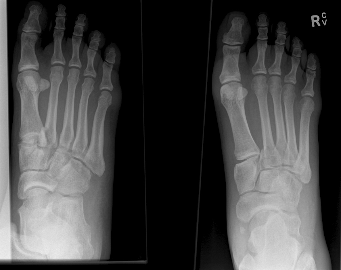
 

Show References


Category: Orthopedics

Title: LisFranc Fractures

Keywords: lisfranc, fracture (PubMed Search)

Posted: 5/17/2014 by Michael Bond, MD (Updated: 4/23/2024)
Click here to contact Michael Bond, MD

Lisfranc Fracture:

Typically consists of a fracture of the base of the second metatarsal and dislocation, though it can also be associated with fractures of a cuboid.  Common current mechanism of injury is when a person steps into a hole and twists the foot.  The original mechanism of injury that was described was when a horseman would fall of their horse with their foot still trapped in a stirrup.

Diagnosis should be considered if patient has difficultly weight bearing with pain on palpation over the 2nd and 3rd metacarpal head with an appropriate mechanism.

Pearls:

  • Fracture findings on plain films may be subtle.
  • If in doubt obtain weight bearing AP views of the foot to demonstrate dislocation/fracture.
  • If weight bearing films are negative and you are still suspicious consider a CT scan of the foot.


Category: Orthopedics

Title: DeQuervains versus Intersection Syndromes

Keywords: DeQuervain, Intersection, Syndrome, Tenosynovitis (PubMed Search)

Posted: 3/30/2014 by Michael Bond, MD (Updated: 4/23/2024)
Click here to contact Michael Bond, MD

DeQuervain and Intersection Syndromes:
 

  • DeQuervain's Syndrome (Tenosynovitis of the Abductor Pollicus Longus and Extensor Pollicus Brevis tendons) is a common disorder that has received a lot of press lately as BlackBerry Thumb or Gamer's Thumb.
    • This condition can be diagnosised by the Finklestein test [Have the patient bend their thumb into the palm of their hand, and then make a fist.  They should then ulnar deviate their wrist.  Pain along the tendons secures the diagnosis.]
    • The pain of DeQuervain's syndrome is typically along the distal end of the radius at the base of the thumb.
  • Intersection syndrome is a less common disorder though closely related to DeQuervain's Syndrome
    • The pain is usually felt on the top of the forearm about three inches proximal to the wrist. 
    • The pain from this condition is due to tenosynovitis of the Extensor carpi radialis longus and Extensor Carpi radialis brevis muscles/tendons caused by the intersection of them with the Extensor pollicus brevis and Abductor pollicus longus tendons.
    • Occurs due to excessive wrist movements.
    • Intersection syndrome can be seen in weight lifters, skiers, and can be seen in homeowners in the fall and winter when they rake a lot of leaves or shovel snow.
  • Treatment is the similar for both conditions and consists of:
    • NSAIDS
    • Cortisone injections can be effective
    • Thumb and wrist immobilization with a Thumb Spica Splint or Cock Up Wrist Splint


Carbon Monoxide is a odorless but deadly gas.  It is important to note that CO has an elimination half-life and it varies under different conditions.
 
When evaluating a patient, we can calculate backwards to determine the COHb level at time of exposure in an acute event.   

Carbon Monoxide Half-Life:

  • Average elimination on room air: 5-6 hours
  • 100% Oxygen: 70-130 minutes
  • 100% Oxygen under hyperbaric conditions at 3 ATA: 23 minutes
There is NO need to recheck COHb level again after initial level because it will be lower- (except in the case of Methylene Chloride exposure).

Show References


Category: Misc

Title: Cause of Post-Operative Fever

Keywords: Postoperative, fever, cause (PubMed Search)

Posted: 1/18/2014 by Michael Bond, MD (Updated: 4/23/2024)
Click here to contact Michael Bond, MD

Post Operative Fever is extremely common, and with the increase in same day surgery this is a common complaint presenting to the ED.  The mnemonic "5Ws" are often taught to remember the causes. They are:

  • Wind - Pneumonia, aspiration, pulmonary embolism, and atelectasis
  • Water - urinary tract infection
  • Walking - Deep Venous Thrombosis or pulmonary embolism
  • Wound - surgical wound infection
  • Wonder drugs - Drug fever, or infection due to indwelling lines, or a reaction to blood products

Though many surgical textbooks report that atelectasis is the most common cause of early post-operative fever,  some even claiming that it is responsible for over 90% of febrile episodes in the first 48 hours after surgery; a recent review in CHEST (reference below) showed that there is no evidence to support this. We often see atelectasis in medical patients too, and few if any of them have fever.  The CHEST review found that there was no clear evidence that atelectasis causes fever at all.

Pearl:  Temperature >38.9C should raise concern for a true infection, where lower temperatures can be due to pulmonary embolism, DVT, drug fever, etc….
 

Show References


"Frozen in January, Amputate in June"  - By Kinjal Sethuraman and Doug Sward
 
Frostbite can lead to major tissue damage even if initial presentation does not look so severe. Treatment is NOT the same as for burns. 

Treatment of Major Frostbite:
1.  Rapid rewarming ASAP of affected area in 40 Celsius degree water until area is thawed (pink and pliable) Logistics are difficult because you have to maintain a constant water temperature- but only if you can maintain same degree of warmth.  Rewarming and refreezing will lead to inevitable tissue death.
2. Wound care, Aloe Vera, ASA 
3.  DELAY surgery except in cases of sepsis or compartment syndrome.
 
CUTTING EDGE:
  • If less than 24 hours since injury, consider diagnostic angiography and  intra-arterial TPA, and heparin infusion, Prostacyclin infusion.
  • Angiography and Bone Scan can be used to prognosticate clinical course.
  • Consider Hyperbaric Oxygen Therapy for moderate to severe frostbite- multiple case reports of significant improvement with HBOT even if delayed by several days. 
 
Treatment of  Minor Frostbite:
1. Rewarm area
2. Ibuprofen
3. Aloe Vera and dressing changes
 
Reference attached from Wilderness Medical Society Practice Guidelines for the Prevention and Treatment of Frostbite  http://www.ncbi.nlm.nih.gov/pubmed/21664561

Show References