UMEM Educational Pearls - By Michael Bond

Category: Orthopedics

Title: Treatment of Back Pain

Keywords: Benzodiazepines, Back Pain, Sciatica (PubMed Search)

Posted: 5/8/2010 by Michael Bond, MD (Updated: 5/9/2010)
Click here to contact Michael Bond, MD

Conservative Treatment of Back Pain:

Muscle relaxanats and benzodiazipnes are often used in the non-operative management of sciatica and non-specific low back pain.  In fact, a 2003 Cochrane review concluded that muslce relaxanats were effective in the management of non-specific low back pain. However, a recent analysis of randomized trials reported little efficacy or only  minor benefits with the use of benzodiazapines in treatment of low back pain.

A recent prospective, randomized, placebo-controlled, double-blinded trial conducted in Germany that enrolled a total of 60 patients found that the use of diazepam was equivilant to placebo in the reduction of distance of referred pain at day 7 of treatment.  Diazepam was also noted on average to increase the length of stay of those patients hospitalized by 2 days (median hospital days of 8 for placebo versus 10 for diazepam), and the probablility of pain reduction on a visual analog scale by more than 50% was twice as high in the placebo group (p< 0.0015).  Placebo reduced the patients pain more than diazepam.

Though the sample size was small; this study should really make one reevaluate the use of diazepam in the treatment of back pain.  Early movement and discouraging bed rest have been associated with decreased back pain, so one mechanism by which  benzodiazepines may make things work is by causing enough sedation to prevent early movement.

Show References


Category: Orthopedics

Title: Carpal Tunnel Syndrome

Posted: 4/25/2010 by Michael Bond, MD (Updated: 3/28/2024)
Click here to contact Michael Bond, MD

Carpal Tunnel Syndrome (CTS):

  • A compressive neuropathy of the median nerve at the wrist as it travels through the carpal tunnel. 
  • Median nerve is bound on three sides by carpal bones and anteriorly by the transverse carpal ligament.  Surgical repair typically consists of cutting this ligament to allow decompression of the nerve.
  • The neuropathy results in:
    • parasethesia of the thumb, index and middle fingers
    • weaknesss of the thumb and thenar muscles.
  • NO physical exam test has great senstivity or specificity for CTS. The two most common are:
    • Phalen's test: hyperflexion of the wrist. Need to hold for 60 seconds.  Sensitivity ~68% and Specificity ~73%
    • Tinel Sign: tapping over cubital tunnel to produce parasthesia along the median nerve. Sensitivity ~50% and Specificity ~77%.
  • Increased risk in those patients with:
    • Diabetes
    • Rheumatoid arthritis
    • hypothyroidism
    • amyloidosis


Category: Ophthamology

Title: Conjunctivitis

Keywords: Conjunctivitis (PubMed Search)

Posted: 4/15/2010 by Michael Bond, MD (Emailed: 4/17/2010) (Updated: 8/28/2014)
Click here to contact Michael Bond, MD

All to often we see children that are sent to the ED for "Pink Eye" as the school nurse will not allow them back into class unless they are treated with antibiotics.  A recent study out of New York identified 4 factors that are associated with low risk (<8% chance) of bacterial (culture postive) conjunctivitis.  They are:

  1. age 6 years
  2. presentation during April through November
  3. watery or no discharge
  4. no glued eye in the morning

An editorial in journal watch comments that if this study can be replicated in other geographic areas we could change the practice of prescribing antibiotics that are not necessary.

Show References


Category: Orthopedics

Title: Prosthetic Hip Dislocatoins

Keywords: Hip Dislocation, Treatment (PubMed Search)

Posted: 4/11/2010 by Michael Bond, MD (Updated: 3/28/2024)
Click here to contact Michael Bond, MD

Prosthetic hip dislocations are a common occurance in the Emergency Department.  After you have gotten the hip back in place there are several ways to prevent the hip from coming out again.  An abductor pillow will work but it confines the patient to bed.  A better option to prevent further hip dislocations until the patient can get an appropriate brace made or reparative surgery is to place the patient in a straight leg knee immoblizer. It is nearly impossible to dislocate your hip if your knee is fully extended.

So after reduction of their simple hip dislocation (i.e: no fractures) place the patient in a straight leg knee immobolizer and they can followup with their orthopedist as an outpatient.


Category: Orthopedics

Title: Ossification Centers of the Elbow in Children

Keywords: Ossification Centers, Elbow (PubMed Search)

Posted: 3/27/2010 by Michael Bond, MD (Updated: 3/28/2024)
Click here to contact Michael Bond, MD

Review of the Appearance of Ossification Centers in Children's Elbows

Determing if a child's elbow has a fracture or if you are looking at an ossification center is easier if you remember the mnemonic CRITOE.  This is the order that the ossification centers appear:

  • Capitellum 1 to 8 months
  • Radial Head 3 to 5 years
  • Internal (medial) Epicondyle 5 to 7 years
  • Trochlea 7 to 9 years
  • Olecranon 8 to 11 years
  • External (Lateral) Epicondyle 11 to 14 yeras


Category: Misc

Title: Critical Care Billing

Keywords: Billing, Critical Care (PubMed Search)

Posted: 3/20/2010 by Michael Bond, MD
Click here to contact Michael Bond, MD

Critical Care billing is time dependent and includes all time spent caring for and coordinating (i.e.: reviewing records, talking to consultants or family) the care of the patient except for the time spent doing separately billable procedures (i.e. central line, CPR, etc).  The following procedures taken from the ACEP website are included in the Critical Care code so the time spent doing these procedures should BE included in your total Critical Care time . 

They are :

  • The interpretation of cardiac output measurements
  • Interpretation of chest x-rays
  • Interpretation of pulse oximetry
  • Interpretation of blood gases, and information data stored in computers
  • Placement of Oral or Nasal gastric tube
  • Temporary transcutaneous pacing
  • Ventilatory management (i.e.: Adjusting the vent, but not the intubation)
  • Vascular access procedures (i.e.: peripherial access)

ACADEMIC MEDICINE CAVEAT: For the reporting of time-based services, such as critical care or moderate sedation, the teaching physician must be directly present during the entire reported time period.

Show References


Category: Orthopedics

Title: Knee Dislocation

Keywords: Knee, Dislocation (PubMed Search)

Posted: 3/13/2010 by Michael Bond, MD (Updated: 3/28/2024)
Click here to contact Michael Bond, MD

Knee Dislocation:

  • It is not uncommon for a patient to have dislocated their knee and it to spontanously reduce prior to presenting to the ED. 
  • Consider the possibility of a spontaneously reduced knee dislocation in any patient with bicruciate (ACL and PCL) ligament instability.  
  • Normal pulses and capillary refill does not exclude occult vascular injury to the popiteal artery.
  • At a minimum the patient should have Ankle Brachial Indexs performed and if <0.9 serial exams and Doppler ultrasound studies should be obtained.
  • Angiography is not absolutely required, and several studies have shown that a selective approach to angiography is acceptable.  As the studies below showed, most patients with findings requiring operative repair on angiography had abnormal physical exams.

Show References


Category: Orthopedics

Title: Pelligrini-Stieda Lesion

Keywords: Pelligrini, Steida (PubMed Search)

Posted: 3/6/2010 by Michael Bond, MD
Click here to contact Michael Bond, MD

Pelligrini-Stieda Lesion:

A Pelligrini-Stieda lesion is shown in the radiograph below.  This lesion was originally described in 1905, and is associated with a tear of the Medial Collateral Ligament.  Heterotrophic calcification forms causing chronic pain, which typically needs to be surgically excised.


So for the students out there, it is possible to diagnosis an MCL tear on plain radiographs.  Just not very often.


Category: Orthopedics

Title: Segond Fracture

Keywords: Segond Fracture (PubMed Search)

Posted: 2/27/2010 by Michael Bond, MD (Updated: 3/28/2024)
Click here to contact Michael Bond, MD

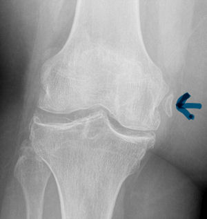
The Segond Fracture:

An benign appearing avulsion fracture of the lateral tibeal plateau that is marker for more significant injuries such as:

  1. Anterior Cruciate Ligament (ACL) tear associated with this fracture 75-100% of the time
  2. Injury to the Medial Meniscus occurs with a Segond fracture 66-75% of the time.

If this avulsion fracture is seen consider immobilzing the patients knee until they can follow up with Orthopedics and/or get an MRI to determine if additional injuries are present.


Category: Orthopedics

Title: Spine CT Scans

Keywords: Spine, Fracture, Diagnosis (PubMed Search)

Posted: 2/20/2010 by Michael Bond, MD
Click here to contact Michael Bond, MD

A recent study by Smith et al showed that the general abdomen/pelvic CT scan in trauma patients obtained with 5mm slices is a better screening test for spine fractures than plain films. They also showed that when compared to dedicated reconstructed thoracolumbar CT scan (2mm slices focused on the spine) it did not miss any clinically significant fractures.

The statistic for plain radiographs and the nonreconstructive CT scan are shown below.

 
Plain Radiographs
Nonreconstructive CT Scan
 
Lumbar
Thoracic
Lumbar
Thoracic
Sensitivity % [95% CI]
47 [33 to 62]
13 [3 to 32]
94 [83 to 99]
73 [50 to 89]
Specificity % [95%  CI]
91 [78 to 97]
71 [54 to 85]
95 [85 to 99]
94 [79 to 99]
Positive Predictive Value % [95% CI]
85 [66 to 96]
15 [2 to 45]
95 [86 to 99]
89 [67 to 99]
Negative Predictive Value % [95% CI]
61 [48 to 72]
56 [41 to 71]
93 [82 to 99]
83 [66 to 93]

The take home point is that dedicated Spine CT scans are probably not needed unless they are going to be used to guide surgical or non-surgical management, and plain films should probably be abandoned in patients that are undergoing CT scans of the chest/abdomen/pelvis.

Show References


Impingement Syndrome and the Diagnostic Accuracy of 5 Common Tests

It is also reported that subacromial impingement syndrome (SAIS) is the more frequent cause of shoulder pain.

The authors of this study attempted to determine the diagnostic accuracy of the following 5 tests for SAIS:

  • Hawkins-Kennedy
  • Neer
  • Empty Can
  • Painful Arc
  • External Resistance

The study demonstrated that any 3 positive tests out of the 5 has a sensitivity of 0.75 (0.54-0.96) , specificity of 0.74 (0.61-0.88), positive likelihood ratio of 2.93 (1.60-5.36) and negative likelihood ratio of 0.34 (0.14-0.80).  See the table below for the individual test characteristics.  No single test was deemed accurate enough to make the diagnosis by itself.


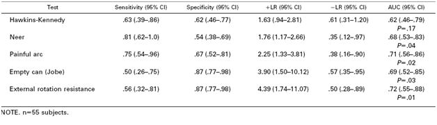
So in the end you should be familiar with most of these tests in order to use a combination of them to make the diagnosis of impingement syndrome.  Future pearls will review how to perform these tests.

Show References


Category: Orthopedics

Title: Scaphoid Fractures

Keywords: Scaphoid, Fracture (PubMed Search)

Posted: 2/6/2010 by Michael Bond, MD
Click here to contact Michael Bond, MD

Scaphoid Fractures:

For suspected scaphoid fractures with negative radiographs it is common practice to put a person in a short arm thumb spica splint until followup up radiographs can be obtained in 10-14 days.

However, there is evidence that a short arm thumb spica splint is not enough for people that have a true scaphoid fracture.  Gellman et al demonstrated that long arm thumb-spica cast immobilization for six weeks followed by short arm thumb-spica cast immobilization decreased time to union by 25% when compared to short arm thumb-spica casting alone.

The theory is that the short arm splint still allows for forearm rotation that can cause shearing motion of the volar radiocarpal ligaments.  A long arm splint prevents this shearing action.  The disadvantage of a long arm splint though is potential elbow joint stiffness and muscle atrophy that can occur during the prolonged period of immobilization.

So for your next patient with a scaphoid fracture seen on radiographs place them in a long arm thumb spica splint.

Show References


Category: Misc

Title: Temporal Arteritis

Keywords: Temporal Arteritis (PubMed Search)

Posted: 1/30/2010 by Michael Bond, MD (Updated: 3/28/2024)
Click here to contact Michael Bond, MD

Temporal Arteritis (TA) is commonly associated with the sudden onset of a unilateral headache centered around the temporal region.  The most devastating consequence of TA is blindness though this is only reported in up to 50% of cases though can be bilateral in up to 33% of patients.

According to the American College of Rheumatology criteria for classification of temporal arteritis this diagnosis can be made in the ED without a biopsy.  You just need at least 3 of the following 5 items to be present (sensitivity 93.5%, specificity 91.2%) to make the diagnosis :

  1. Age of onset older than 50 years
  2. New-onset headache or localized head pain
  3. Temporal artery tenderness to palpation or reduced pulsation
  4. Erythrocyte sedimentation rate (ESR) greater than 50 mm/h
  5. Abnormal arterial biopsy (necrotizing vasculitis with granulomatous proliferation and infiltration)

Show References


Category: Airway Management

Title: Uveitis (Cont'd)

Keywords: Uveitis, Treatment (PubMed Search)

Posted: 1/23/2010 by Michael Bond, MD (Updated: 3/28/2024)
Click here to contact Michael Bond, MD

Uveitis and Iritis Treatment:

  • Once the diagnosis is suspected or made ensure that the patient has ophthamology followup.
  • Antibiotics are not needed as this is not an infectious process.
  • Pain control is the painstay of therapy (no not narcoletics) but cycloplegics like:
    • Cyclopentolate 0.5-2% 1 gtt TID
    • Homatropine 2-5% 1 gtt TID
    • This will relieve pain and photophobia symptoms
  • Topical steroid can be initiated to decrease inflammation but should be done in consultation with the ophthamologist
    • Prednisolone 1% 1 gtt every 1-6 hours.


Category: Ophthamology

Title: Uveitis and Iritis

Keywords: Uveitis, Iritis (PubMed Search)

Posted: 1/16/2010 by Michael Bond, MD
Click here to contact Michael Bond, MD

Iritis is a common diagnosis in the ED, but did you know it was actually a subset of Uveitis.

Uveitis is an inflammation of one or all parts of the uveal tract which consists of the iris, the ciliary body, and the choroid.
 
The subsets of uveitis are:

  1. anterior
  2. confined to the iris and anterior chamber -- iritis
  3. confined to the iris, anterior chamber, and ciliary body -- iridocyclitis.
  4. Posterior uveitis -- choroiditis and chorioretinitis, is uncommon, with the exception of cytomegalovirus (CMV) retinitis in patients with AIDS.


Treatment of iritis and uveitis next week.

Show References


Category: Orthopedics

Title: Paronychia

Keywords: Paronychia (PubMed Search)

Posted: 1/9/2010 by Michael Bond, MD
Click here to contact Michael Bond, MD

Acute paronychia

  • Usually result from minor trauma of the skin around the fingernail such as biting, manicures, picking a hangnail or finger sucking.
  • Staphylococcus aureus is the most common infecting organism. However other mouth flora such as Streptococcus and Pseudomonas species, gram-negative bacteria, and anaerobic bacteria can also be a cause.
  • Recommended treatement consists of incision and drainage and placing the patient on  amoxicillin /  clavulanic acid or clindamycin to cover all the organisms noted above.


Category: Dermatology

Title: Pityriasis Rosea

Keywords: Pityriasis rosea (PubMed Search)

Posted: 1/3/2010 by Michael Bond, MD
Click here to contact Michael Bond, MD

Pityriasis Rosea

  • A common exantham that typically presents initially with a herald patch, followed by a generalized rash over the next 1-2 weeks and can last 4-6 weeks.
  • Patients may initially have prodromal symptoms such as joint pain,headache, fever and malaise that precede the appearance of the rash.
  • The primary plaque, herald patch, is seen on the skin in 50-90% of cases.
  • The generalized rash typically develops in crops along the lines in the skin leading to a characteristic "Christmas tree" pattern.
  • Pruritus is present in 75% of cases.
  • Usually a self-limited, benign illness that does not require any treatment.  Though symptomatic treatment of the pruritus is reasonable.


Category: Dermatology

Title: Nikolsky's sign

Keywords: Nikolsky's sign, Dermatology (PubMed Search)

Posted: 12/26/2009 by Michael Bond, MD (Updated: 3/28/2024)
Click here to contact Michael Bond, MD

Nikolsky's sign is positive when slight rubbing of the skin results in exfoliation of the skin's outermost layer.  The more technical term is acantholysis which is the loss of the normal adhesion of the epithelial skin cells which allows for this sloughing.

Seen in:

  • Toxic Epidermal Necrolysis
  • Pemphigus vulgaris
  • Scalled Skin Syndrome
  • Bullous impetigo
  • Epidermolysis bullosa

Often helpful to differentiate pemphigus vulgaris from bullous pemphigoid. The sign is usually absent in bullous pemphigoid.  Just be careful with how much testing you are doing as this can be very painful to the patient.


Category: Misc

Title: Hypothermia

Keywords: Hypothermia (PubMed Search)

Posted: 12/19/2009 by Michael Bond, MD (Updated: 3/28/2024)
Click here to contact Michael Bond, MD

Hypothermia Pearls:

  • Lidocaine is generally ineffective in preventing ventricular arrhythmias, as is cardiac pacing or atropine to increase the heart rate.
  • Should the patient fully arrest be prepared to perform CPR for a long time.  If your ED does not have a automatic CPR device consider calling your local fire department or ambulance service as they might have one that can be loaned to your department.
  • Warm fluids, heated blankets and heat lamps will typically increase a patients temperature about 1' C an hour.
  • Gastric lavage, peritoneal lavage and heated IV fluids can warm as much as 3' an hour.
  • To rewarm quickly as high as 18'C an hour requires cardiac bypass or thoracic lavage.

Finally, remember to monitor the patient closely when you first start rewarming as this can induce cardiac arrest.  This is thought to occur as colder peripherial blood returns to the central circulation as peripherial veins and arteries dilated from the warm fluid.


Category: Airway Management

Title: Patella Fractures

Keywords: Patella, Fracture (PubMed Search)

Posted: 12/13/2009 by Michael Bond, MD (Updated: 3/28/2024)
Click here to contact Michael Bond, MD

Patella fractures are typically due to direct trauma as in a fall or direct blow to the knee.

Fractures may be missed on the AP view or misdiagnosed as a bipartate fracture.  To avoid these pitfalls look closely at the lateral view and consider getting a sunrise view of the knee (better visualizes the patella).  Finally,  unilateral bipartate patella are very rare so consider an x-ray of the contralateral knee if you are considering this as your diagnosis.

Surgery should be considered for:

  • Fractures with displacement greater than 3 mm.
  • Individuals that have lost there externsor mechanism as it is indicative of a tear in the extensor retinacula.