Category: Airway Management
Keywords: Ketamine, etomidate, RSI, induction (PubMed Search)
Posted: 12/19/2024 by Robert Flint, MD
(Updated: 6/17/2026)
Click here to contact Robert Flint, MD
Another large database evaluation of the use of etomidate vs. ketamine as an induction agent for intubation found a trend toward higher mortality in the etomidate group. Even when trying to control for steroid use (to control for etomidate’s possible adrenal suppression), etomidate had a higher mortality rate.
A well done study that adds to the chorus advocating for choosing ketamine when looking for a hemodynamically neutral induction agent.
Wunsch H, Bosch NA, Law AC, Vail EA, Hua M, Shen BH, Lindenauer PK, Juurlink DN, Walkey AJ, Gershengorn HB. Evaluation of Etomidate Use and Association with Mortality Compared with Ketamine among Critically Ill Patients. Am J Respir Crit Care Med. 2024 Nov 15;210(10):1243-1251. doi: 10.1164/rccm.202404-0813OC. PMID: 39173173.
Category: Airway Management
Keywords: major adverse event, airway, management, cardiovascular collapse (PubMed Search)
Posted: 7/30/2023 by Robert Flint, MD
(Updated: 6/17/2026)
Click here to contact Robert Flint, MD
This systemic review and meta analysis looked at major adverses events (hypoxia, cardiovascular instability, or cardiac arrest) in patients intubated in emergency departments, ICU’s, or medical floors. They found nearly 1/3 of patents had an event. ICU intubation and patients with pre-existing hemodynamic compromise had the highest rate of adverse outcomes. This study gives further support to the concept of maximizing resuscitation pre-intubation and to anticipate a major event peri-intubation. Be prepared and don't be surprised when something doesn't go as planned.
Category: Airway Management
Keywords: hypotension, pharmacology, RSI (PubMed Search)
Posted: 6/9/2023 by Robert Flint, MD
(Updated: 6/17/2026)
Click here to contact Robert Flint, MD
Take away: Be prepared (with blood products and/or vasopressors) for hypotension in trauma patients post-intubation particularly the elderly and severely injured. Pre-intubation tachycardia predicts post-intubation hypotension. Resuscitation with saline in traumatically injured patients is inferior to blood products or permissive hypotension.
A UK study retrospectively looked at trauma patients undergoing helicopter based emergency medicine intubation using induction agents of fentanyl, ketamine, and rocuronium for hypotensive episodes. “This study demonstrates that more than one in five patients who undergo PHEA have a new episode of significant hypotension within the first ten minutes of induction. Increasing patient age, multi-system injuries, a higher baseline heart rate, and intravenous crystalloid administration by the ambulance service before HEMS arrival were all significantly associated with PIH, whereas the addition of fentanyl to the induction drug regime was not.”
Price, J., Moncur, L., Lachowycz, K. et al. Predictors of post-intubation hypotension in trauma patients following prehospital emergency anaesthesia: a multi-centre observational study. Scand J Trauma Resusc Emerg Med 31, 26 (2023). https://doi.org/10.1186/s13049-023-01091-z
Category: Airway Management
Keywords: leg pain, compartment syndrome (PubMed Search)
Posted: 10/9/2022 by Brian Corwell, MD
(Updated: 6/17/2026)
Click here to contact Brian Corwell, MD
Popliteal artery entrapment syndrome (PAES)
CC: Exertional lower leg pain, however, compression of posterior neurovascular structures can lead to nonspecific vascular and neurogenic symptoms.
Challenging diagnosis to make because of close overlap with chronic exertional compartment syndrome (CECS).
Anatomic PAES has a prevalence of 0.62% to 3.5% in the general population. Patients are more likely to be older be older, male, and have lower levels of activity.
Functional popliteal artery entrapment (FPAE) however has no anatomic anomaly. Sx’s are thought to be because of bulky surrounding muscle crowding with repetitive dynamic injury. This is most commonly from the medial head of the gastrocnemius. Patients are younger and more likely to be involved in athletics. Most athletes were involved in sports that put high value on repetitive plantarflexion, such as track and field (45%), soccer (25%), water sports (8%), lacrosse (6%), basketball (6%),
Sx’s: bilateral (25-75% of cases) cramping in the region of the soleus and plantar paresthesias.
Common exacerbating mechanism: ascending stairs or climbing inclines because of leg/knee position of extension with plantarflexion
In one review, 31% of patients who underwent debulking surgery for FPAES had been previously treated and extensively worked up at outside institutions for CECS, and already undergone various compartment releases.
Patients in one study underwent a dynamic CTA protocol. A positive test demonstrated normal flow in neutral position and compression or complete occlusion of the popliteal artery by the medial head of the gastrocnemius muscle against the lateral femoral condyle with provocative foot plantarflexion. Images below.
https://images.journals.lww.com/acsm-csmr/Original.00149619-202210000-00008.F1.jpeg
Nearly three-fourths of athletes limited by FPAES demonstrated full return to prior competitive levels with four compartment fasciotomy AND surgical debulking of the anterolateral quadrant of the medial head of the gastrocnemius muscle.
Lawley RJ,et al., Concurrent Diagnosis of Functional Popliteal Artery Entrapment Syndrome and Chronic Exertional Compartment Syndrome in Athletes. Curr Sports Med Rep. 2022 Oct 1;21(10):366-370.
Category: Airway Management
Keywords: trauma, PTX, finger thoracostomy, needle decompression, 2nd intercostal space, 5th intercostal space, pneumothorax (PubMed Search)
Posted: 9/25/2022 by Robert Flint, MD
(Updated: 6/17/2026)
Click here to contact Robert Flint, MD
Finger thoracostomy is superior to needle decompression in the fifth mid-axiallary intercostal space which is superior to the traditionally taught needle decompression in the second mid-clavicular intercostal space for traumatic tension pneumothorax/trauamtic arrest.
SHARON HENRY, MD, FACS ATLS 10th edition offers new insights into managing trauma patients Bulletin of the American College of Surgeons PUBLISHED JUNE 1, 2018
Scott Weingart, MD FCCM EMCRIT Podcast 62 – Needle vs. Knife II: Needle Thoracostomy? December 11, 2011
Hannon, L. et al. .Finger thoracostomy in patients with chest trauma performed by paramedics on a helicopter emergency medical service Emerg Med Australas 2020 Aug;32(4):650-656.doi: 10.1111/1742-6723.13549. Epub 2020 Jun 21
Andy Neil Stop putting IV cannulae in the 2nd ICS for tension PTX Emergency Medicine Ireland Posted on November 15, 2012
Category: Airway Management
Keywords: knee pain, running injury (PubMed Search)
Posted: 9/24/2022 by Brian Corwell, MD
Click here to contact Brian Corwell, MD
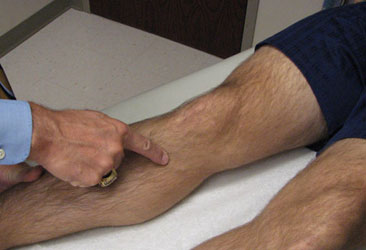
Pes Anserinus pain syndrome (formerly pes anserine Bursitis)
Occurs at the bursa of the pes anserinus which overlies the attachment of the 1) Sartorius 2) gracilis and 3) semi-tendinosis tendons. Insertions resemble a Goose’s foot.
An inflammatory condition of the medial knee
Location is 2-3 inches below the knee joint on the medial side
1st layer of medial compartment
https://www.dramynrajani.com/wp-content/uploads/2018/05/pes-anserine-bursitis-clinical-test.jpg
Patients complain of knee pain just below medial joint line (esp with stairs)
History may include sudden increase in running distance especially with hills (common)
Associated with obesity, tight hamstring muscles and with knee OA
PE: Tenderness to palpation of the bursa possibly with mild swelling
DDx: MCL tear, medial meniscus injury, medial (knee) compartment arthritis, tibial stress fracture
Treatment: Cessation/modification of offending activities, Icing and ice massage, NSAIDs, hamstring stretching and physical therapy. Failure of the above should prompt referral for bursal steroid injection.
Category: Airway Management
Keywords: Concussion, risk, head impact (PubMed Search)
Posted: 6/11/2022 by Brian Corwell, MD
(Updated: 6/17/2026)
Click here to contact Brian Corwell, MD
Head Impact Exposure and Concussion Incidence
There has been a major focus on head impact biomechanics as a cause of single-impact concussion in football.
The role of repeated subclinical (without diagnosed concussion) head impact exposure (HIE)
during the preseason and regular season may also be contributory.
There may exist individualized concussion tolerance levels. This threshold may be reduced by the burden of sustained subconcussive impacts
NCAA Division 1 football athletes sustain a median of 426 impacts over the course of a football season
652 impacts/season in high school football
Total head impact exposure during the preseason occurred at 2x the rate of the regular season
This association was investigated over 1120 athlete seasons from 6 NCAA D1 football programs across 5 years
Head Impact Telemetry was used to record head impact exposure
Elevated preseason HIE was strongly associated with preseason and in season concussion incidence
Total season HIE was strongly associated with total season concussion incidence.
Conclusion: There is a prolonged effect of HIE on concussion risk starting with preseason football.
Athletes with higher preseason HIE may have higher risk of concussion for the entire fall season.
In Practice:
In 2016, the Ivy League eliminated full contact practices from the regular season in addition to their existing limits on the amount of full contact in practice during the spring and preseason.
Currently, the NCAA has the following limitations: Teams won’t be allowed to hold full-contact practices on more than two days in a row. Each practice session is limited to only 75 minutes of full contact, in addition to a limit of two preseason scrimmages.
Stemper BD, et al; CARE Consortium Investigators. Association between Preseason/Regular Season Head Impact Exposure and Concussion Incidence in NCAA Football. Med Sci Sports Exerc. 2022 Jun 1;54(6):912-922.
Category: Airway Management
Keywords: Hemorrhage, Pre-hospital, Trauma, Shock (PubMed Search)
Posted: 6/9/2022 by Lucas Sjeklocha, MD
Click here to contact Lucas Sjeklocha, MD
Enthusiasm for early transfusion of blood products in patients with traumatic shock has increased with increasing availability of pre-hospital blood and plasma and results of studies such as the PAMPer trial of pre-hospital plasma have shown potential mortality benefits. The deployment of prehospital blood for patients in hemorrhagic shock is promising but has significant cost and logistical considerations.
The RePHILL trial was a UK pre-hospital-based study of packed red blood cells and lyophilized plasma versus normal saline in trauma patients with presumed hemorrhagic shock. Patients older than age 16 with an SBP<90 or an absent radial pulse were eligible to get up to 1L of the study intervention. Multiple centers took part in the trial with 1:1 randomization stratified by study center. The primary outcome was a combination of mortality or lactate clearance less than 20% per hour or both.
A total of 432 patients were assigned a study fluid. The population was 82% male, median of 38 years old, with 78% of injuries classified as blunt, and 82% of the presumed hemorrhage classified at non-compressible. This was a very ill population with an average SBP of 73, an average GCS of 7 and an ISS of 36. The average from emergency call to EMS arrival was 30 minutes, average to study intervention was 26 minutes and time from EMS activation to ED arrival was 90 minutes.
The results showed no difference in the primary composite endpoint (64% vs 65%), with no difference in mortality (43% vs 45%) or lactate clearance (50% vs 55%). Interestingly, patients in the blood product arm had similar vital signs, lactate, and INR on ED arrival but received more blood products in the first 24 hours after ED arrival (pRBC 6.34 vs 4.41, p=0.004 and Plasma 5.04 vs 3.37, p=0.002). The was a trend toward improved early mortality at 3hr in the pre-hospital blood group (16% vs 22%, p=0.08).
Bottom Line(s):
Prehospital packed red blood cells and lyophilized plasma as compared to saline for traumatic shock did not improve mortality or lactate clearance in a well conducted multicenter RCT.
The use of prehospital blood products is promising but population which benefits, and the optimal type of product and delivery mechanism remain unclear.
Increased blood utilization and lower early mortality in the blood product group may represent alteration in the spectrum of disease that requires different early management.
The reasons for this counterintuitive result are unclear and further trials of whole blood as well as fibrinogen concentrates are ongoing.
Resuscitation with blood products in patients with trauma-related haemorrhagic shock receiving prehospital care (RePHILL): a multicentre, open-label, randomised, controlled, phase 3 trial. Crombie et al. Lancet Hematology. 2022.
https://doi.org/10.1016/ S2352-3026(22)00040-0
Category: Airway Management
Keywords: PRP, hematoma, muscle tear (PubMed Search)
Posted: 3/12/2022 by Brian Corwell, MD
Click here to contact Brian Corwell, MD
Treatment of Hamstring Strains in Athletes
28 year old athlete presents to the ED and diagnosed with a hamstring strain
Localized swelling, moderate pain and a small limp. Incomplete tearing of the muscle
He is worried that he will miss the remainder of his season and when he returns will reinjure the same hamstring
Consider referral to sports medicine/orthopedics
A recent study looked at use of ultrasound guided hematoma aspiration followed by platelet-rich plasma (PRP) treatment on recovery in acute hamstring injuries
55 male athletes between ages 18 -32 weighing between 170 and 260lbs
27 with treatment protocol plus rehabilitation and 28 treated conservatively (rehabilitation)
All had Grade 2 hamstring injuries diagnosed on MRI
Partial muscle tear (<50% cross sectional area)
Note: Grade 2 hamstring injuries are often associated with INTERmuscle hematoma and subsequent scarring. This can lead to persistent pain/discomfort and reinjury
Average return to play time was 32.4 days in the standard of care group
Average return to play time was 23.5 days in the intervention group (P<0.001)
Recurrence rate of hamstring strain was 28.6% in the standard of care group
Recurrence rater of hamstring strain was <4% in the intervention group (P=0.025)
Athletes with grade 2 hamstring injuries treated with hematoma aspiration and PRP injection into the strain had significantly shorter return-to-play and much lower recurrence rate that athletes treated with rehabilitation alone
Trunz LM, et al. Effectiveness of Hematoma Aspiration and Platelet-rich Plasma Muscle Injections for the Treatment of Hamstring Strains in Athletes. Med Sci Sports Exerc. 2022 Jan 1;54(1):12-17.
Category: Airway Management
Keywords: Caffeine, Exercise, VO2 max (PubMed Search)
Posted: 12/25/2021 by Brian Corwell, MD
Click here to contact Brian Corwell, MD
Caffeine is probably the most wildly used and studied drug/supplement in the world.
It has been shown to enhance exercise capacity and performance.
Mechanism of action is likely multifactorial and involves adenosine receptor antagonism via direct CNS action improving mental alertness, reaction time and reducing the perceived exertion rate (pain).
To no surprise, amateur and elite athletes use caffeine to improve performance.
The well-accepted dosage of caffeine to improve performance is between 3 and 6 mg/kg, approximately 60 min before exercise. This dosage promotes (between 1 and 8%) performance gains in aerobic exercises and exercises with high glycolytic demand from cyclists to tennis players to weightlifters.
Consider the lower end of this range if interested in trying this on your own.
In an evaluation of 20,686 urine samples of elite athletes, almost 75% of the samples contained caffeine in concentrations higher than 0.1 μg/mL
Caffeine also increases maximal oxygen uptake (VO2 max)
23 elite athletes were tested twice with and twice without caffeine.
Randomized, double-blinded, placebo-controlled study.
Caffeine 4.5 mg/kg taken 45 minutes before exercise
Measures: Time to exhaustion and VO2 max.
Caffeine increased time to exhaustion and VO2 max, thereby increasing overall performance.
If you are going to incorporate using caffeine before your next workout, I suggest espresso shots for extra caffeine without the volume of a large cup of coffee. Beware of known side effects such as jitters, anxiousness and difficulties with sleep if taken later in the day. Also consider stomach upset digestive issues, and increased heart rate.
Happy Holidays!!!!
Category: Airway Management
Keywords: Myocarditis, Covid-19 (PubMed Search)
Posted: 1/23/2021 by Brian Corwell, MD
(Updated: 6/17/2026)
Click here to contact Brian Corwell, MD
Exercise and Covid-19
The majority of COVID-19 cases fall into the mild-to-moderate category, with symptoms lasting less than 6 weeks on average.
The disease presents a challenge for clinicians seeking to offer counsel for patients wishing to return to exercise.
A recent cohort study in Germany looked at 100 patients (avg. age 49, 53% male) who had recovered from Covid-19 infection.
Most had been healthy, with no pre-existing medical conditions, before becoming infected.
The group had cardiac MRI (CMR) performed.
Average time interval between Covid-19 diagnosis and CMR was 71 days.
Cardiac involvement was seen in 78% of patients and ongoing myocardial inflammation in 60%.
Evidence based return to activity guidelines being developed are more conservative than in the past with other viral infections
https://link.springer.com/article/10.1007/s11420-020-09777-1/tables/1
1) Puntmann VO, Carerj ML, Wieters I, et al. Outcomes of Cardiovascular Magnetic Resonance Imaging in Patients Recently Recovered From Coronavirus Disease 2019 (COVID-19). JAMA Cardiol. 2020;5(11):1265–1273. doi:10.1001/jamacardio.2020.3557
2) Metzl, J.D., McElheny, K., Robinson, J.N. et al. Considerations for Return to Exercise Following Mild-to-Moderate COVID-19 in the Recreational Athlete. HSS Jrnl 16, 102–107 (2020).
Category: Airway Management
Keywords: Patient, centered, communication (PubMed Search)
Posted: 5/30/2020 by Michael Bond, MD
(Updated: 6/17/2026)
Click here to contact Michael Bond, MD
Asking these allows everybody to understand what the goal really is — what are you really fighting for? It’s for a life that contains certain things.
Gawande, A. (2014). Being mortal : medicine and what matters in the end / Atul Gawande: Medicine and what matters in the end / Atul Gawande (First edition.). New York: Metropolitan Books : Henry Holt & Company.
Category: Airway Management
Keywords: Epidural abscess, back pain (PubMed Search)
Posted: 3/14/2020 by Brian Corwell, MD
(Updated: 6/17/2026)
Click here to contact Brian Corwell, MD
Laboratory studies are not often indicated in the early evaluation of low back pain.
Complete blood counts (CBC) have poor sensitivity and specificity for infection. White blood cell (WBC) counts, have poor sensitivity and specificity for infection. They may be elevated and a left shift or bandemia may be present and increase suspicion for infection, but a lack of these does not rule out infection. Elevated WBC counts are only found in two-thirds of patients with SEA.
Both erythrocyte sedimentation rate (ESR) and C-reactive protein (CRP) are highly sensitive (84-100%) for spinal infections and are observed in >80% with vertebral osteomyelitis and epidural abscesses. However, elevated CRP was found in 87% of patients with an epidural abscess as well as half of patients with spine pain not due to an epidural abscess, so is not highly specific.
CRP levels rise rapidly and decrease rapidly with improvement in disease and may be better used to follow response to treatment. ESR is the most sensitive and specific serum marker of infection. ESR is elevated in 94-100% of patients with an epidural abscess compared to only 33% of those without an epidural abscess. Infection is unlikely in patients with an ESR less than 20 mm/h. Although an elevated ESR (>20 mm/h) is the most specific serum test for infection, it also may indicate occult malignancy (sensitivity, 78%; specificity, 67%).
If infection is suspected, obtain two sets of blood cultures, as a causative pathogen may be identified in ~50% of patients.
Category: Airway Management
Keywords: MRI, back pain (PubMed Search)
Posted: 2/22/2020 by Brian Corwell, MD
Click here to contact Brian Corwell, MD
Cauda Equina Syndrome is a medical emergency that is considered in all patients who present to the ED with lower back pain.
Clinical presentation is variable in nature and may include some combination of lower back pain, bowel or bladder dysfunction, sexual dysfunction, saddle anesthesia with motor/sensory abnormalities.
MRI is the gold standard for diagnosis
Many of us have encountered a scenario where a patient with high clinical suspicion returns with scan negative MRI.
Studies have attempted to characterize this population.
Patients in the scan negative group had an increased prevalence of functional disorders (37% vs. 9%), functional neurologic disorders (12% vs. 0%), and psychiatric comorbidities (53% vs. 20%).
Further study is needed to characterize this association.
Hospitals may consider individualized neurologic and psychiatric referral for certain patients who are scan negative in the future.
Is scan-negative cauda equina syndrome a functional neurological disorder? A pilot study. Gibson et al., Eur J Neurol 2020, Feb 19.
Is scan-negative cauda equina syndrome a functional neurological disorder? A pilot study. Gibson et al., Eur J Neurol 2020, Feb 19.
Category: Airway Management
Keywords: back pain, urinary retention, CES (PubMed Search)
Posted: 1/11/2020 by Brian Corwell, MD
(Updated: 6/17/2026)
Click here to contact Brian Corwell, MD
Known effects and side effects of prescribed medicines may masquerade as cauda equina syndrome (CES) .
Analgesic medicines used by patients with chronic back pain may also cloud the diagnosis of CES.
Cholinergic medications (glaucoma/myasthenia) may lead to voiding issues.
Anticholinergic medications (COPD/urinary incontinence) may lead to urinary retention.
Opioids – Constipation, reduced bladder sensation
Anticonvulsants (Gabapentin/Pregabalin)- Urinary incontinence
Antidepressants (Amitriptyline) – Urinary retention, sexual dysfunction, reduced awareness of need to pass urine
NSAIDs – Urinary retention.
Verhamme KM, et al. Nonsteroidal anti-inflammatory drugs and increased risk of acute urinary retention. Arch Intern Med. 2005:165;1547-1551.
Category: Airway Management
Keywords: Adrenal Crisis (PubMed Search)
Posted: 1/7/2020 by Caleb Chan, MD
(Updated: 6/17/2026)
Click here to contact Caleb Chan, MD
Adequate treatment of adrenal crisis (AC) is often delayed, even when a h/o adrenal insufficiency is known.
Besides refractory hypotension, also consider in pts with:
Beware of triggers:
Treatment:
Amrein K, Martucci G, Hahner S. Understanding adrenal crisis. Intensive Care Med. 2018;44(5):652-655.
Rushworth RL, Torpy DJ, Falhammar H. Adrenal Crisis. N Engl J Med. 2019;381(9):852-861.
Category: Airway Management
Keywords: HLH, Hemophagocytic Lymphohistiocytosis (PubMed Search)
Posted: 12/24/2019 by Kim Boswell, MD
Click here to contact Kim Boswell, MD
Hemophagocytic Lymphohistiocytosis (HLH) – Part I
A rare, but important disease that is becoming more widely recognized and more frequently diagnosed. This disease, while uncommon, is rapidly progressive and caries a high mortality rate.
Causes are not completely understood, but involve abnormal activation of the immune response due to a failure of the typical downregulation in hyperinflammatory processes.
Two types exist:
Congenital/Familial – genetic predisposition which usually requires a triggering event to occur
Acquired – occurs in adults with no known predisposition (often have underlying genetic predispositions) – triggering events include infections , immunodeficiency, rheumatologic disorders, and malignancy in addition to many others.
Diagnosis is challenging due to the wide variety of symptoms and constellation of symptoms, which often mimic more common infections/sepsis presentations. Common symptoms include the following:
Symptoms can, and do, occur in any body system – rashes, conjunctivitis, DIC, LFT abnormalities, hypotension/shock, and respiratory failure are all common concomitant findings in the presentation of HLH
More on the specific diagnosis and treatment to follow in part II...
McClain KL. Clinical features and diagnosis of hemophagoctyic lymphohistiocytosis. UpToDate.Waltham, MA:UpToDate Inc. https://www.uptodate.com (Accessed on December 24, 2019.)
Category: Airway Management
Keywords: PE, tachypnea, Critical Care, ED Disposition (PubMed Search)
Posted: 10/21/2019 by Robert Brown, MD
Click here to contact Robert Brown, MD
ICU admission rates for all acute PEs vary wildly across the country (<5% to ~80%).
To predict which hemodynamically stable, normotensive PE patients should be admitted to the ICU, a single-center retrospective analysis of 7 years’ data sought to describe the reasons why normotensive patients with PE required vasopressors within 48 hours of admission to the ICU. The authors studied 293 patients admitted to the ICU at Beth Israel Deaconess in Boston and found only 8 patients (2.7%) who decompensated within the first 2 days. Of MANY variables studied, only respiratory rate was significantly different between those who decompensated and those who did not (mean RR 29 with range 26-32 in the decompensated group vs mean 21 with range 17-24).
Bottom Line: cost control experts may lean on you to admit fewer PE patients to the ICU. There is no perfectly reliable way to predict which normotensive patient with a PE will decompensate. The PESI score has been validated but even the low risk cohort had 1.6% mortality at 3 days. The BOVA score has been validated but its endpoint of mortality at 30 days is less useful for planning admission. Tachypnea should concern you.
Admon A, Seymour C, Gershengorn H, et al. Hospital-level variation in ICU admission and critical care procedures for patients hospitalized for pulmonary embolism. Chest 2014; 146(6):1452-1461
Patel H, Shih J, Gardner R, et al. Hemodynamic decompensation in normotensive patients admitted to the ICU with pulmonary embolism. Journal of Critical Care 2019; 54:105-109
Category: Airway Management
Keywords: prothrombin complex concentrate, warfarin, bleeding (PubMed Search)
Posted: 5/29/2019 by Ashley Martinelli
(Updated: 6/1/2019)
Click here to contact Ashley Martinelli
For patients with bleeding due to warfarin, prothrombin complex concentrate (PCC) is the recommended antidote. Historically, PCC has been dosed on weight and INR:
· INR 2 - 4: 25 units/kg, max 2500 units
· INR 4 - 6: 35 units/kg, max 3500 units
· INR > 6: 50 units/kg, max 5000 units
New data demonstrates that fixed dosing offers several advantages with similar efficacy outcomes:
· Standardized dosing
· Improved time to administration
· Decreased cost
The University of Maryland Health System has adopted a fixed dose strategy for all patients with warfarin-associated critical bleeding:
· Bleeding site other than intracranial hemorrhage AND INR 1.4 - 6 AND weight ≤ 100 kg = 1500 units
· Intracranial hemorrhage OR > 100 kg OR INR >6 = 2000 units
**Note: PCC is also the antidote of choice for reversing critical bleeding due to factor Xa inhibitors (rivaroxaban, apixaban, edoxaban). All critical bleeds due to these agents should receive 50 units/kg, max 5000 units.
Klein L, Peters J, Miner J, Gorlin J. Evaluation of fixed dose 4-factor prothrombin complex concentrate for emergent warfarin reversal. Am J Emerg Med. 2015;33: 1213-12128.
Astrup G, Sarangarm P, Burnett A. Fixed dose 4-factor prothrombin complex concentrate for the emergent reversal of warfarin: a retrospective analysis. J Thrombosis Thrombolysis. 2018;45:300-305.
Scott R, Kersten B, Baisor J, Nadler M. Evaluation of fixed-dose four-factor prothrombin complex concentrate for emergent warfarin reversal in patients with intracranial hemorrhage. J Emerg Med. 2018;54(6):861-866.
Category: Airway Management
Keywords: had, wrist, carpal (PubMed Search)
Posted: 1/26/2019 by Brian Corwell, MD
(Updated: 6/17/2026)
Click here to contact Brian Corwell, MD
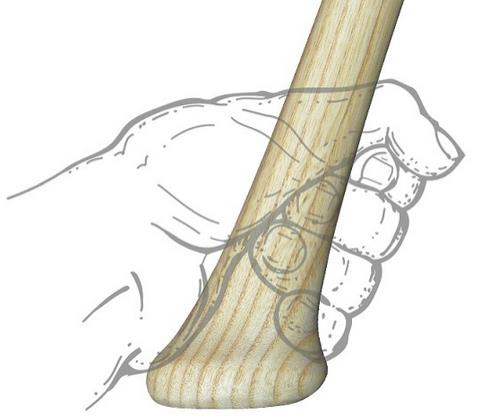
Hook of hamate fracture
Often missed fracture despite classic history
A frequent athletic injury
Seen in stick sports (golf, baseball, hockey)
Typically caused by a direct blow (grounding a gold club)
https://upload.orthobullets.com/topic/6035/images/hamate_baseball.jpg
Patient presents with hypothenar pain and pain with tight gripping
https://upload.orthobullets.com/topic/6035/images/hamate_golf.jpg
Presentation may be subacute with longstanding wrist or palmer pain
Physical exam: Tender to palpation over hook of hamate
Specialized test: hook of hamate pull test
Supinated hand held in ulnar deviation. Ask patient to actively flex 4th and 5th digits against resistance at DIP.
https://www.youtube.com/watch?v=A-mjRnC1yWQ
Radiology: Consider adding carpal tunnel view to standard wrist series if diagnosis is suspected
CT sometimes needed to image the fracture
Tx: Immobilize in a short arm splint
https://eorthopod.com/news/new-test-for-sports-injury-of-the-hand/
https://www.orthobullets.com/hand/6035/hook-of-hamate-fracture