As a follow up to Dr. Winter’s Pearl on Anaphylaxis on 1/24/2017, here’s a handy pearl for pediatric anaphylaxis (part 1).
Anaphylaxis: rapid and potentially life-threatening involvement of at least 2 systems following exposure to an antigen.
Medications (max: adult doses)
Get it?!?! Easy right? Instead of fumbling through an app or reference card during your next case of pediatric anaphylaxis, be a rock star "EM DR" by remembering the “Rule of 2’s”.
(Can't help it...ya'll know I love my mnemonics!!)
More studies are needed, but the existing data shows that medical adhesives may be quicker without impacting cosmetic and functional outcome.
In pediatrics, providers typically prescribe 10 mg/kg (max 500 mg) and 5 mg/kg daily x 4 (max 250 mg) for treatment of pneumonia, but this dosing regimen is NOT recommended for all azithromycin usage. There are other dosing regimens that are important to keep in mind during the respiratory season:
1) Pharyngitis/ tonsillitis (ages 2-15 yr): 12 mg/kg daily x 5 days (max 500 mg/ 24 hr)
2) Pertussis
3) Acute sinusitis >/= 6 months: 10 mg/kg daily x 3 days
Which first-line vasoactive drug is the best choice for children with fluid-refractory septic shock? A prospective, randomized, blinded study of 120 children compared dopamine versus epinephrine in attempts to answer this debated question in the current guidelines for pediatric sepsis.
Bottom line: Dopamine was associated with an increased risk of death and healthcare–associated infection. Early administration of peripheral or intraosseous epinephrine was associated with increased survival in this population.
Using 1.5 mg/kg or 2 mg/kg of IV ketamine led to less redosing compared to using 1 mg/kg IV.
Typically, empiric treatment for lobar community acquire pneumonia (CAP) in immunized < 5 year olds (preschool) is amoxicillin (45mg/kg BID or 30 mg/kg TID for resistant S. pneumoniae) for outpatient and ampicillin or ceftriaxone for inpatient. Additional coverage with azithromycin is typically recommended for school age and adolescent patients (>= 5 years), but not necessarily for younger children unless there is a particular clinical suspicion for atypical pneumonia with history, xray findings, or sick contacts.
However, in sickle cell patient with suspicion for acute chest syndrome, azithromycin is recommended for all ages groups, as atypical bacteria such as Mycoplasma are a common cause of acute chest syndrome in patients of all ages with sickle cell disease even young children. In a prospective series of 598 children with acute chest syndrome, 12% of the 112 cases in children less than 5 had positive serologic testing of M. pneumoniae (9% of all cases had M. pneumoniae) (Neumayr et al, 2003).
Plasma-Lyte A outperformed 0.9% NaCl for rehydration in children with acute gastroenteritis showing a more rapid improvement in serum bicarbonate levels and dehydration scores.
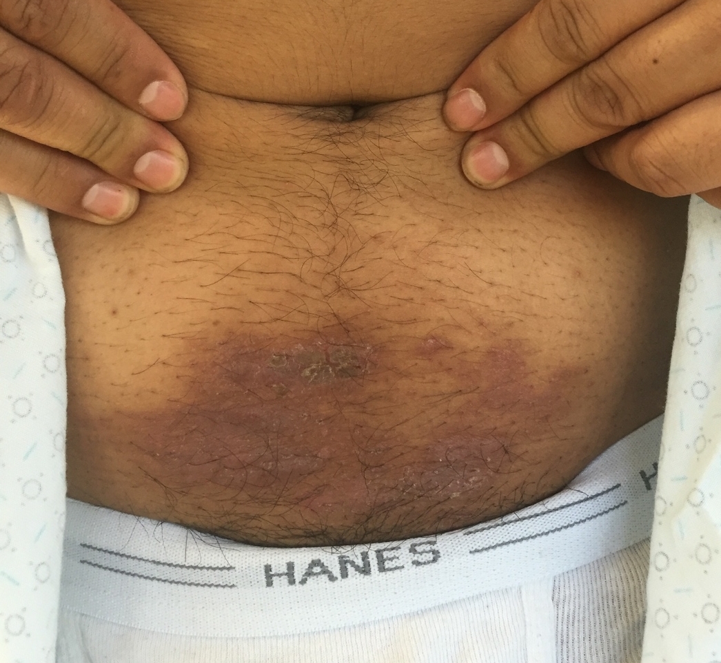
A 12 year old male who recently started middle school presents to the ED with a rash in the periumbilical region that has been developing over the last few weeks. The rash is scaly, somewhat itchy, but otherwise benign appearing. The patient has no known medical conditions other than eczema, and is otherwise well. What is the diagnosis?
Picture courtesy of Mara Haseltine, MD
114 children with bronchiolitis had end tidal carbon dioxide (ETCO2) measured on presentation to the ED. The ETCO2 levels did not differ significantly between admitted and discharged patients. In the subset of admitted patients, there was no correlation with ETCO2 on admission and days of oxygen requirement or length of stay.
Bottom line: Initial ETCO2 does not predict outcome for patients with bronchiolitis.
From 2010-2014 ED visits in the US for injuries from trampoline parks (TPI) increased from 581 visits per year to 6932 visits per year. There was no change in the number of injuries related to home trampoline use. TPI were more likely to involve the lower extremity, be a dislocation and warrant admission and less likely to involve the head.
Bottom line: TPIs are increasing and have a different injury pattern compared to home trampolines.
The pediatric epiglottis is more "U" shaped, often overlies the glottic opening, and is "less in line with the trachea."1 Because of this, it has traditionally been taught that a Miller blade is the ideal laryngoscope.
Varghese et al compared the efficacy of the Macintosh blade and the Miller blade when placed in the vallecula of children between the ages of 1 and 24 months. The blades provided similar views and suffered similar failure rates. When the opposite blade was used as a backup, it had a similar success rate as the opposing blade.2 Passi et al also compared these two blades, this time placing the Miller blade over the epiglottis. Again, similar views were achieved.3
Although it is summer, preparations are being made for the 2016-2017 influenza season. The Center for Disease Control (CDC) no longer recommends the live attenuated influenza vaccine (LAIV4). The American Academy of Pediatrics has supported this statement.
The LAIV4 (the only intranasal vaccine available) was offered to patients over the age of 2 years without respiratory problems. Observational studies during the 2013-2015 seasons have shown that the LAIV4 has an adjusted vaccine efficacy of 3% compared to 63% for the inactivated vaccine (intramuscular). Children who received the intranasal vaccine were almost 4 times more likely to get the flu compared to children who received the injection.
Bottom line: Only the intramuscular shot is recommended for this upcoming season. This is causing many primary care practices to scramble to obtain enough vaccine.
The American Academy of Pediatrics has developed a new set of clinical practice guidelines to help better manage and think about patients who have experienced an ALTE (Apparent Life Threatening Event). The term BRUE (Brief Resolved Unexplained Event) will replace ALTE.
BRUE is defined as an event in a child younger than 1 year where the observer reports a sudden, brief and now resolved episode of one or more of: cyanosis or pallor; absent, decreased or irregular breathing, marked change in tone or altered level of responsiveness. A BRUE can be diagnosed after a history and physical exam that reveal no explanation.
BRUE can be classified as low risk or high risk. Those that can be categorized as low risk do not require the extensive inpatient evaluation that has often occurred with ALTE.
LOW risk BRUE:
Age > 60 days
Gestational age at least 32 weeks and postconceptual age of at least 45 weeks
First BRUE
Duration < 1 minute
No CPR required by a trained medical provider
No concerning historical features (outlined in the article)
No concerning physical exam findings (outlined in the article)
Recommendations for low risk BRUE:
-SHOULD: Educate, shared decision making, ensure follow up and offer resources for CPR training
-May: Obtain pertussis and 12 lead; briefly monitor patients with continuous pulse oximetry and serial observations
-SHOULD NOT: Obtain WBC, blood culture, CSF studies, BMP, ammonia, blood gas, amino acids, acylcarnitine, CXR, echocardiogram, EEG, initiate home cardiorespiratory monitoring, prescribe acid suppression or anti-epileptic drugs
-NEED NOT: obtain viral respiratory tests, urinalysis, glucose, serum bicarbonate, hemoglobin or neuroimaging, admit to the hospital solely for cardiorespiratory monitoring
*When looking at the evidence strength behind these recommendations, the only one that had a strong level was that you should not obtain WBC, blood culture or CSF
Neonatal jaundice- Incidence ~85% of term newborns
Bili levels are EXPECTED to rise during first 5 days of life
Be aware of CONJUGATED hyperbilirubinemias (biliary atresia, infection)
Majority of cases due to increase in unconjugated (indirect) bilirubin 2/2 residual fHgb breakdown and insufficient capacity of hepatic conjugation
Severe hyperbilirubinemia (Tbili >20mg/dL) <2% of term infants
⇒
Acute bilirubin encephalopathy(ABE)- Hypertonia, arching, opisthotonos, fever, high pitched cry
⇒
Kernicterus (5% of ABE)-CP, MR, auditory dysfunction, upward gaze palsy
When to refer for phototherapy/exchange transfusion
Typically, if an infant or young child presents to the ED with concern for intracranial hemorrhage (ICH), CT is performed as a rapid diagnostic tool. Now that clinicians are more aware of the radiation associated with head CT, the possible use of ultrasound was studied. Ultrasound is commonly used in the neonatal population for detecting ICH. A study by Elkhunovich et al looked at children younger than 2 years who had cranial ultrasounds preformed. Over a 5 year period, 283 ultrasounds were done on patients between 0 to 485 days old (median 33 days). There were 39 bleeds detected. Ultrasound specificity and sensitivity was calculated by comparing the results with CT, MRI and/or clinical outcome. For significant bleeds, the sensitivity for ultrasound was 81%. The specificity for detecting ICH was 97%.
Only 2 patients in the study were older than 1 year. The proper windows are easiest to visualize in children younger than 6 months.
Bottom Line: The sensitivity of cranial ultrasound is inadequate to justify its use as a screening tool for detection of ICH in an infant with acute trauma, but it could be considered in situations when obtaining advanced imaging is not an option because of availability or patient condition.
A previous pearl has looked at serum HCO3 as a predictor of DKA (see pearl from 8/21/15). The article by Gilhotra looks at using end tidal CO2 (ETCO2) to exclude DKA. 58 pediatric patients were enrolled with 15 being in DKA. No patient with ETCO2 > 30 mmHg had DKA. Six patients with ETCO2 < 30 mmHg did not have DKA. Other studies done in children have shown similar results.
An article recently published by Chebl and colleagues examined patients older than 17 years with hyperglycemia. In this study, 71 patients were included with 32 having DKA. A ETCO2 >35 excluded DKA in this group while a level <22 was 100% specific for DKA.
Bottom line: ETCO2 >35 mmHg is a quick bedside test that can aid in the evaluation of hyperglycemic patients.
Perianal Group A Strep is an infectious dermatitis seen in the perianal region that is caused by Group A beta-hemolytic Strep. Children will have a characteristic rash with a sharply-demarcated area of redness, swelling, and irritation around the perianal region. There may be associated swelling and irritation of the vulva and vagina (in girls) and penis in boys. Patients can have bleeding or itching during bowel movements.
The age range is often <10 years of age. There is often an absence of fever or other systemic symptoms.The diagnosis can be confirmed by obtaining a Rapid Strep swab from the area of interest. You can also collect a bacterial culture of the area.
Treatment requires a 14 day course of penicillin. Amoxicillin (40 mg/kg/day divided TID) and clarithromycin are alternative treatments. The additional of topical bactroban (mupirocin) can be effective, but it should not be used as monotherapy. Re-occurrence is common, so close follow-up is key.