UMEM Educational Pearls - By WanTsu Wendy Chang

Category: Neurology

Title: Neurological Adverse Reactions with Antimicrobials

Keywords: drug reaction, toxicity, neurotoxicity, antibiotics (PubMed Search)

Posted: 11/10/2021 by WanTsu Wendy Chang, MD (Emailed: 11/11/2021)
Click here to contact WanTsu Wendy Chang, MD

  • Antimicrobial medications can be associated with neurological adverse reactions. 
  • An individual’s risk is influenced by their age, weight, nutritional status, the medications they are taking concurrently, and pharmacological properties (dosage, half-life, CNS permeability). 
  • Encephalopathy 
    • Seen with beta-lactams, fluoroquinolones, clarithromycin, and sulfamethoxazole-trimethoprim. 
    • Most commonly with cefepime. 
    • Higher risk in elderly, renal dysfunction, and preexisting CNS disease. 
  • Seizures 
    • Beta-lactams block GABA receptors. 
    • Highest risk with cefepime and imipenem. 
  • Peripheral neuropathy 
    • Associated with metronidazole, fluoroquinolones, linezolid, chloramphenicol, and isoniazid. 
    • Most cases are dose dependent. 
    • Some cases are irreversible. 
  • Ototoxicity 
    • Aminoglycosides cause cochlear NMDA receptor excitotoxicity. 
  • Weakness 
    • Fluoroquinolones, macrolides, and aminoglycosides inhibit acetylcholine release and bind neuromuscular junction receptors. 
    • Should be avoided in myasthenia gravis and Lambert-Eaton syndrome. 
  • Movement disorders 
    • Tremors – sulfamethoxazole-trimethoprim 
    • Dyskinesia, dystonic reactions – fluoroquinolones, chloramphenicol 
    • Cerebellar syndrome – metronidazole, aminoglycosides 

Bottom Line: Recognition of antibiotic associated neurotoxicity reduces unnecessary workup and serious adverse effects. 

Show References


Category: Neurology

Title: Emergency Department Burr Hole (Submitted by Dr. Christina Powell)

Keywords: burr hole, trephination, subdural hematoma, epidural hematoma, herniation (PubMed Search)

Posted: 10/13/2021 by WanTsu Wendy Chang, MD
Click here to contact WanTsu Wendy Chang, MD

Your patient presents with a large traumatic subdural hematoma with midline shift and clinical evidence of herniation.  Your nearest neurosurgeon is several hours away, what do you do?

Initial resuscitation should follow ATLS.  Treatment of intracranial hypertension and herniation includes elevating the head of bed, administering osmotic therapies, optimizing analgesia/sedation, and hyperventilation.  If all measures have been exhausted and there is a delay to definitive neurosurgical intervention, an emergency department burr hole may be considered.

Indications:

  • GCS < 8, dilated and nonreactive pupil(s), posturing suggestive of uncal or transtentorial herniation 
  • Radiographic evidence of an extra-axial (subdural/epidural) hematoma causing midline shift and brainstem compression
  • Lack of timely neurosurgical intervention
  • Procedure will not delay transfer to definitive care

Contraindications:

  • Neurosurgical intervention available within reasonable time frame
  • Skull fracture at site of planned burr hole

Equipment:

  • Razor
  • Surgical marker
  • Sterile prep and drape
  • Syringe, needle, lidocaine
  • Scalpel, forceps, retractor, sharp hook, scissors
  • Hand drill, hex wrench, drill bit with guard
  • Sterile saline, gauze, dressing

Transtemporal Approach:

  • Measure skull thickness on CT for depth of drill guard.
  • Position patient supine and elevate the ipsilateral shoulder with a shoulder roll.  Utilize tape or have assistant hold the head in place. 
  • Shave the hair.
  • Mark the point 2 cm superior and 2 cm anterior to the tragus.
  • Sterile prep and drape.
  • Inject local anesthetic and then make a 3 cm vertical skin incision down to the periosteum.  Dissect and use a retractor to expose the skull.
  • Drill with steady pressure perpendicular to the skull.  Irrigate with sterile saline to remove bone fragments.
  • Once the skull is penetrated:
    • If an epidural hematoma, blood should be released.  Can use sterile saline to facilitate drainage of clotted blood.
    • If a subdural hematoma, use a sharp hook to tent the dura and make a small cruciate incision.
  • Place loose sterile dressing.
  • Transfer to definitive care.

Additional Points:

  • Neurosurgery consultation before performing this procedure is recommended. 
  • Antibiotic prophylaxis with gram-positive coverage is recommended.
  • In extenuating circumstances, this may be considered without CT confirmation of the location of the extra-axial hematoma.  However, there is risk of a negative exploratory burr hole due to a hematoma not in the temporal location or due to a false localizing sign.

Show References


Category: Neurology

Title: Thrombectomy for Basilar Artery Occlusion?

Keywords: stroke, large vessel occlusion, basilar artery, posterior circulation, thrombectomy (PubMed Search)

Posted: 6/9/2021 by WanTsu Wendy Chang, MD
Click here to contact WanTsu Wendy Chang, MD

  • The landscape of acute ischemic stroke treatment changed dramatically with endovascular thrombectomy (EVT).
  • However, few patients with basilar artery occlusions were included in major EVT trials.
  • Basilar artery occlusion accounts for 10% of large vessel occlusions and can result in devastating neurological deficits.
  • The recently published BASICS trial evaluated the efficacy of EVT within 6 hours of symptom onset in 300 patients with basilar artery occlusion strokes.
  • 44.2% of the EVT group had a good outcome compared to 37.7% of the medical treatment group (p=0.19).
    • Good outcome was defined as modified Rankin scale of 0 (no symptoms) to 3 (moderate disability but able to walk without assistance) at 90 days.
    • Symptomatic intracranial hemorrhage was higher in the EVT group (4.5% vs. 0.7%, p=0.06).
    • History of AFib was more common in the EVT group (28.6% vs. 15.1%).
  • It is important to note that this study did not use advanced neuroimaging for patient selection unlike in landmark EVT trials of anterior circulation large vessel occlusion strokes.

Bottom Line: There is no significant difference between endovascular thrombectomy and medical management for basilar artery occlusion strokes within 6 hours of symptom onset. 

Show References


Category: Neurology

Title: Factors that Decrease Post-Lumbar Puncture Headaches

Keywords: Lumbar puncture, LP, post-dural, headache, intracranial hypotension (PubMed Search)

Posted: 5/12/2021 by WanTsu Wendy Chang, MD (Updated: 3/19/2024)
Click here to contact WanTsu Wendy Chang, MD

  • Post-lumbar puncture (LP) headache, reported in up to 33% of patients, is due to a persistent CSF leak causing intracranial hypotension.
  • A recent review by Cognat et al. looked to answer several frequently asked clinical questions:
    • Who is at decreased risk of post-LP headache?
      • Infants and children have a similar prevalence compared to adults.
      • Older patients have a lower risk, with an incidence of <5% in those over 60 years old.
    • Does needle choice minimize the risk of post-LP headache?
      • Atraumatic non-cutting ("Whiteacre" or "Sprotte") needles have lower rates (RR 0.4, 0.34-0.47).
      • The use of atraumatic needles does not affect the rate of success, success on first attempt, or duration of the LP.
    • Does performing the LP in a specific way prevent post-LP headache?
      • LPs performed in the lateral decubitus position and at a higher intervertebral space have a lower incidence.
      • Difficult LPs (e.g. multiple attempts, traumatic tap) do not appear to affect the rate.
      • The volume of CSF removed does not affect the rate.
    • Do any treatments after the LP reduce post-LP headache occurrence?
      • Bed rest after LP does not reduce and may in fact worsen the likelihood.
      • Fluids and caffeine do not prevent post-LP headaches.

Bottom Line: The use of atraumatic needles is most effective in reducing the risk of post-LP headaches. These needles are easy to use and have similar rate of success as cutting needles.

Show References


Category: Neurology

Title: Functional Neurological Disorders in the ED

Keywords: functional neurological disorder, FND, stroke mimic, non-epileptic seizure (PubMed Search)

Posted: 4/28/2021 by WanTsu Wendy Chang, MD
Click here to contact WanTsu Wendy Chang, MD

  • Functional neurological disorders (FND) are unintentional and involuntary. 
  • Imaging and electrophysiological studies have shown cerebral dysfunctions in attention and perception, which may explain why symptoms often improve with distraction. 
  • Diagnosis requires demonstration of inconsistency and/or incongruency with recognized neurological or medical conditions. 
    • No clinical sign alone is diagnostic. 
    • Patients may have comorbidities such as multiple sclerosis, stroke, or epilepsy. 
  • Hoover’s sign and drift without pronation have been described as positive signs for FND. 
    • These can also be seen in patients with pain, neglect, or apraxia. 
  • Avoid maneuvers that may harm the patient, such as dropping their arm onto their face. 
    • A high-pitched tuning fork applied to the nostrils is an effective stimulus to assess responsiveness.  
  • Avoid using terms like non-organic, psychogenic, or pseudoseizure. 
  • When counseling a patient, avoid only explaining what conditions they do not have or attributing symptoms to psychological problems or stress. 
    • Instead, name the diagnosis, explain that their symptoms are real and common, and emphasize that symptoms are potentially reversible. 
  • Early diagnosis of FND is associated with improved physical and psychological outcomes. 

Bottom Line: Functional neurological disorders (FND) are commonly encountered in the ED. A thorough neurological exam may reveal positive signs suggestive of FND. Early diagnosis and referral to specialists may improve outcomes. 

Show References


Category: Neurology

Title: Alteplase Administration Errors in Stroke

Keywords: acute ischemic stroke, alteplase, tPA, thrombolysis, error (PubMed Search)

Posted: 4/15/2021 by WanTsu Wendy Chang, MD
Click here to contact WanTsu Wendy Chang, MD

  • Alteplase administration in acute ischemic stroke (AIS) has been associated with errors.
  • A recent study looked at the types of errors that occurred at regional hospitals and Comprehensive Stroke Centers.
  • 19.8% (133/676) of patients had an error associated with alteplase administration with the majority occurring at regional hospitals without stroke certification.
    • The most common error was over-dosage of alteplase, occurring in 5% (36) of patients.
    • Other common errors included under-dosage, infusion errors, and apparent contraindications.
  • The most common contributing factor leading to the error was incorrect calculation.
  • Administration error was associated with higher rate of hemorrhagic conversion (12.7% vs. 7.1%, p=0.04).

Bottom Line: Alteplase administration in acute ischemic stroke is associated with errors, most commonly with over-dosage of the medication.

Show References


Category: Neurology

Title: The Rising Incidence of Idiopathic Intracranial Hypertension

Keywords: Idiopathic intracranial hypertension, IIH, pseudotumor cerebri, obesity, healthcare utilization (PubMed Search)

Posted: 3/10/2021 by WanTsu Wendy Chang, MD
Click here to contact WanTsu Wendy Chang, MD

  • Idiopathic intracranial hypertension (IIH) predominantly affects women of childbearing age and is strongly associated with obesity.
  • Patients can have high rates of healthcare utilization due to their chronic disabling headaches, multiple diagnostic tests and specialist consultations, and potential complications related to treatments.
  • A recent study looked at trends in the incidence, prevalence, and healthcare outcomes of IIH in the 3.1 million Welsh population over a 14-year period.
    • Incidence of IIH increased from 2.3/100,000 to 7.8/100,000 (p<0.001).
    • Prevalence of IIH increased from 12/100,000 to 76/100,000 (p<0.001).
    • Obesity (BMI >30 kg/m2) also increased from 29% to 40% of the population (p<0.001), with IIH incidence and prevalence strongly associated with BMI.
    • Outcomes included blindness (0.78%), moderate visual impairment (1.9%), CSF diversion (9%) with 44% of these patients requiring at least 1 revision surgery.
    • IIH patients also had 5.28 times higher rate of unscheduled hospital visits during this time period.

Bottom Line: The incidence and prevalence of IIH is increasing, likely related to rising rate of obesity. This has also been associated with more healthcare utilization compared to the general population.


Show References


Category: Neurology

Title: Is There a Seasonal Variation to Bell's Palsy?

Keywords: Bell's palsy, facial palsy, Lyme disease, Borrelia burgdorferi (PubMed Search)

Posted: 2/24/2021 by WanTsu Wendy Chang, MD (Updated: 3/19/2024)
Click here to contact WanTsu Wendy Chang, MD

  • Bell’s palsy can be caused by herpes simplex virus-1 (HIV-1) and Lyme borreliosis.
  • Prior studies have found higher incidence of Bell’s palsy in colder months possibly related to reactivation of latent HSV-1.
  • A recent study looked at the incidence and seasonal variation of positive Lyme tests in Bell’s palsy patients in a Lyme-endemic area (New Jersey).
    • Over a 5-year period, 81% of 442 patients with Bell’s palsy were tested for Lyme and 16% tested positive.
    • The months May through October had a 7.2 times higher incidence of positive Lyme tests in Bell’s palsy patients, with the peak in July.
    • May through October also had a 1.3-fold increased ED visits for Bell’s palsy, also peaking in July.

Bottom Line: In a Lyme-endemic area, the incidence of positive Lyme tests and Bell’s palsy are highest in the Lyme months. This seasonal variation may help guide the management of patients with Bell’s palsy.


Show References


Category: Neurology

Title: Does My Seizure Patient Need An EEG?

Keywords: seizure, status epilepticus, nonconvulsive, electrographic, EEG (PubMed Search)

Posted: 1/27/2021 by WanTsu Wendy Chang, MD (Updated: 3/19/2024)
Click here to contact WanTsu Wendy Chang, MD

  • Most seizures resolve spontaneously, however, seizures that persist >5 minutes or recur without the patient returning to their baseline should be treated expeditiously with benzodiazepines and antiepileptic medications.
  • A subset of patients may continue to have electrographic seizures despite cessation of their convulsive seizure activity.
    • Prior studies described 26-52% of patients develop nonconvulsive seizures after resolution of convulsive status epilepticus.
  • The recent Established Status Epilepticus Treatment Trial (ESETT) compared fosphenytoin, levetiracetam, and valproic acid in aborting seizures and improving responsiveness in patients who did not response to initial treatment with benzodiazepines.
    • EEG was not required for this trial, but 58% (278/478) had an EEG within 24 hours after seizure onset.
      • Of those who had an EEG, 14% (39/278) had electrographic seizures.
    • For patients who had clinical treatment success, 13% (13/102) were found to have electrographic seizures.

Bottom Line: Persistent or recurrent seizures are not uncommon in the first 24 hours after status epilepticus even in patients with resolved clinical seizure activity. Early use of EEG can help identify patients who need further escalation of treatment.


Show References


Category: Neurology

Title: Do All Post-tPA Patients Require ICU Care?

Keywords: stroke, thrombolysis, tissue plasminogen activator, tPA, monitoring (PubMed Search)

Posted: 1/13/2021 by WanTsu Wendy Chang, MD
Click here to contact WanTsu Wendy Chang, MD

  • Acute ischemic stroke patients are commonly admitted to an ICU after receiving IV tPA to be closely monitored for potential complications.
  • Current post-tPA protocol requires frequent vital signs and neurological assessments up to every 15 minutes, thereby requiring 1:1 or 1:2 nurse-to-patient ratio.
  • Studies have shown that stroke severity is a strong predictor of the need for critical care interventions for post-tPA patients, where patients with an NIHSS score ≥10 have an ~8x higher odds of requiring critical care interventions than those with NIHSS <10.
  • A low-intensity post-tPA monitoring protocol (Figure 1) for patients with NIHSS <10 and no critical care needs has been found to be safe in a small single center study.
    • These low risk patients were admitted with telemetry monitoring and 1:3 nurse-to-patient ratio after an initial period of q15 min standard monitoring.
    • An international, multicenter, randomized controlled trial is being planned to study this further.

  • This strategy may help streamline care and utilize hospital resources more efficiently in the COVID-19 pandemic and beyond.

Bottom Line: Patients with NIHSS <10 may be safe for low-intensity post-tPA monitoring if they do not require critical care after an initial period of q15 min standard monitoring for the first 2 hours.

Show References


Category: Neurology

Title: Does Language Preference Affect Acute Stroke Care?

Keywords: acute ischemic stroke, guideline, metric, English, non-English (PubMed Search)

Posted: 12/9/2020 by WanTsu Wendy Chang, MD (Emailed: 12/10/2020) (Updated: 12/10/2020)
Click here to contact WanTsu Wendy Chang, MD

  • Prior studies have shown that ethnic minorities have lower levels of stroke knowledge and lower penetrance of public health stroke education.
  • A recent study looked at whether patients’ language preference affects acute ischemic stroke care metrics.
    • 3190 stroke patients at an urban Comprehensive Stroke Center, where 300 (9.4%) had a non-English preferred language
    • They found no difference in:
      • Time from symptom discovery to ED arrival (128 min vs. 161 min for patients with English preferred language, p=0.68)
      • Arrival by EMS (65% vs. 61.3%, p=0.21)
      • Door-to-imaging time (55 min vs. 60 min, p=0.33)
      • Door-to-needle time for thrombolysis (51 min vs. 53 min, p=0.69)

Bottom Line: Patients' language preference does not appear to affect the efficiency of acute ischemic stroke care, especially at experienced high volume stroke centers. 

Show References


Category: Neurology

Title: What Makes a Headache a Migraine?

Keywords: migraine, headache, diagnosis, treatment, prevention (PubMed Search)

Posted: 11/11/2020 by WanTsu Wendy Chang, MD (Updated: 11/13/2020)
Click here to contact WanTsu Wendy Chang, MD

  • Migraine is the 2nd most common neurologic disorder after tension headache and accounts for more disability than all other neurologic disorders combined.
  • Diagnosis is clinical and defined by at least 5 episodes of headache that meet the following criteria:
    • Duration of 4 to 72 hours (when untreated or unsuccessfully treated)
    • At least 2 characteristics: unilateral, pulsating, moderate-to-severe pain intensity, aggravated by physical activity
    • Accompanied by at least 1 symptom: nausea, vomiting, photophobia, phonophobia
  • Aura symptoms must be fully reversible and may be visual, sensory, speech/language, motor, brainstem, or retinal.
  • Early treatment while the headache is still mild include NSAIDs followed by triptans.
    • Opioids and barbiturates are not recommended due to adverse effects and risk of dependency.
  • Preventive treatment is recommended for patients who have at least 2 migraine days per month and whose lives are adversely affected.
    • Common therapies include antihypertensive agents (e.g. propranolol), antidepressants (e.g. amitriptyline), anticonvulsants (e.g. topiramate, valproate), and calcium-channel blockers (flunarizine).

Bottom Line: Migraine is a common and debilitating condition that benefits from early treatment. Consider initiating preventive therapy for patients who experience at least 2 migraine days per month and adverse effects despite treatment.

Show References


  • When dealing with weakness and sensory complaints in the ED, the time course of symptoms and physical exam findings help identify emergent conditions.
  • We often talk about upper motor neuron vs. lower motor neuron signs that distinguish whether a lesion is in the central or peripheral nervous system.
  • Characteristics that differentiate between central vs. peripheral nervous system pathology include:
  Central Nervous System Peripheral Nervous System
Pattern of Symptoms
Hemibody involvement
Weakness of UE extensors
Weakness of LE flexors
Distal involvement in polyneuropathy
Distal and proximal involvement in polyradiculoneuropathy
Proximal involvement in polyradiculopathy
Sensory often precedes motor symptoms 
Pure proximal>distal weakness may be due to myopathy or NMJ disorder
Sensory Symptoms
• Central poststroke pain (hyperalgesia, allodynia)
• Sensory level in spinal cord pathology 
• Proprioception involved early in dorsal column disorders
Neuropathic pain (burning, tingling, shock-like) 
Ascending sensory loss involving distal BLE>BUE in polyneuropathy
Proprioception involved late in polyneuropathy
Reflexes
Hyperreflexia in affected limb(s) after acute period
Positive Babinski’s sign
Hyporeflexia in affected limb(s)
Tone Increased after acute period Decreased
UE = upper extremity
LE = lower extremity
NMJ = neuromuscular junction
 
Bottom Line: A systematic approach to the evaluation of weakness and sensory complaints in the ED help differentiate between central vs. peripheral nervous system pathology.


Show References


Category: Neurology

Title: CVT Presentation and Management

Keywords: cerebral venous thrombosis, CVT, symptoms, treatment, endovascular (PubMed Search)

Posted: 9/23/2020 by WanTsu Wendy Chang, MD
Click here to contact WanTsu Wendy Chang, MD

  • We've talked about the rising incidence of cerebral venous thrombosis (CVT) and choice of neuroimaging studies before, now let’s talk about presentation and treatment.
  • Symptoms range from headache to coma with cerebral edema and intracranial hypertension depending on the veins and sinuses involved.
    • Superior sagittal sinus is most frequently affected (62%) and can cause headache, hemiparesis, hemisensory loss, hemianopia, and seizures.
    • Transverse sinus is also commonly involved (45%) and can cause headache, aphasia, and seizures.
    • Thrombosis of the deep veins is seen in 18% of cases and can cause altered mental status, coma, and gaze palsy.
  • Management includes anticoagulation, treatment of underlying cause, seizures, and intracranial hypertension.
    • LMWH is preferred unless in patients with renal dysfunction or need for rapid reversal of anticoagulation.
    • Endovascular intervention may be considered in severe cases that do not improve or deteriorate despite anticoagulation.
  • Poor prognostic factors are: 
    • 2 points each - malignancy, coma, deep venous thrombosis
    • 1 point each - mental status disturbances, male, intracranial hemorrhage
    • Score ≥3 suggests high risk of poor outcome

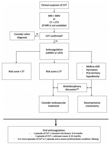
Bottom Line: Severity of CVT presentation depends on the location and clot burden. Anticoagulation is key, though consider endovascular intervention if patient does not improve or deteriorates despite anticoagulation.


Show References


Category: Neurology

Title: The Rising Incidence of Cerebral Venous Thrombosis

Keywords: cerebral venous thrombosis, CVT, prothrombotic, headache (PubMed Search)

Posted: 9/10/2020 by WanTsu Wendy Chang, MD
Click here to contact WanTsu Wendy Chang, MD

  • Cerebral venous thrombosis (CVT) is thought to predominantly affect young and middle-aged females. 
    • Known risk factors include prothrombotic states such as malignancy and oral contraceptive use, as well as local infections and head trauma.
  • The incidence of stroke in young adults is rising worldwide.
  • A recent study by Otite et al. examined the incidence of CVT during 2006-2016 in New York and Florida utilizing the State Inpatient Database.
    • CVT remains an uncommon condition though number of admissions increased 70%.
    • Mean age of patients increased with number of hospitalizations in the elderly doubled.
    • Incidence was highest in Blacks, followed by non-Hispanic Whites and Hispanics.
  • This rise in incidence may be related to increased recognition, improved diagnostic studies, increased neuroimaging utilization, emerging or unknown risk factors. 

Bottom Line: The incidence of CVT is increasing with rate of increase higher in males and older females.  Consider CVT beyond traditional risk factors. 


Show References


Category: Neurology

Title: The Painful Twitch - Trigeminal Neuralgia

Keywords: trigeminal neuralgia, TN, tic douloureux, neuropathic facial pain (PubMed Search)

Posted: 8/26/2020 by WanTsu Wendy Chang, MD
Click here to contact WanTsu Wendy Chang, MD

  • Trigeminal neuralgia is diagnosed by:
    • Pain in 1 or more divisions of the trigeminal nerve
    • Paroxysms of pain that are sudden, intense, usually few seconds in duration
    • Pain triggered by innocuous stimuli in the trigeminal nerve territory (91-99% patients)
  • 24-49% of patients experience continuous or long-lasting pain
  • Exam may reveal forceful contraction of the facial muscles during a paroxysm (tic convulsif)
  • Causes include:
    • Intracranial vascular compression of the trigeminal nerve root (most common)
    • Multiple sclerosis, cerebellopontine angle tumor
    • Idiopathic (10% of cases)
  • Carbamazepine and oxcarbazepine are first-line treatments
    • They may be poorly tolerated due to side effects including dizziness, diplopia, ataxia, CNS depression, and hyponatremia
    • They also have limited efficacy on continuous pain
  • Acute exacerbations may warrant admission for hydration, acute pain control, and titration of antiepileptic drugs
    • Botulinum toxin A was recently added as a treatment option

Bottom Line: New onset trigeminal neuralgia needs workup for its etiology. Carbamazepine and oxcarbazepine can be effective for symptom management though continuous or long-lasting pain exacerbations are difficult to treat.

Show References


Category: Neurology

Title: Is That a CSF Leak?

Keywords: cerebrospinal fluid, rhinorrhea, otorrhea, halo, double ring, beta-2 transferrin (PubMed Search)

Posted: 8/12/2020 by WanTsu Wendy Chang, MD (Updated: 3/19/2024)
Click here to contact WanTsu Wendy Chang, MD

  • Spontaneous cerebrospinal fluid (CSF) rhinorrhea is rare and usually related to a combination of thinning of the bone and dura and fluctuating intracranial pressure.
  • CSF rhinorrhea can be associated with idiopathic intracranial hypertension, skull base tumors, neurosurgical and otolaryngology procedures, and trauma.
  • Trauma with fracture of the anterior skull base is the most common cause of CSF rhinorrhea.
  • CT and MRI can identify bony defects, whereas cisternography can diagnose occult leaks.
  • Fluid containing CSF is classically described to make a “halo” or “double-ring” pattern on gauze or linen.

  • However, this sign is not specific to CSF, as mixtures of blood with saline, tears, or rhinorrhea can also produce halos.
  • Beta-2 transferrin is a protein found almost exclusively in CSF* thus can be used to diagnose CSF rhinorrhea.

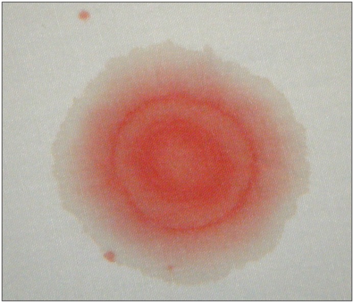
Bottom Line: Beta-2 transferrin is more accurate than the halo sign to identify CSF containing fluid.

Beta-2 transferrin is found in low concentrations in the perilymph in the cochlea, and aqueous and vitreous humor of the eye


Show References


Category: Neurology

Title: The Headache Formerly Known as Pseudotumor Cerebri (Submitted by Ryan Spangler)

Keywords: idiopathic intracranial hypertension, papilledema, intracranial pressure, cranial nerve palsy (PubMed Search)

Posted: 7/8/2020 by WanTsu Wendy Chang, MD
Click here to contact WanTsu Wendy Chang, MD

Question

Idiopathic intracranial hypertension (IIH) is a vision-threatening illness with significant morbidity and needs to be considered as a possible headache diagnosis in the ED. Most often, this occurs in women of childbearing age with a BMI >30, but atypical varieties exist.

Symptoms: Headache (90%), visual disturbance, pulsatile tinnitus, horizotal diplopia.

Signs: Papilledema, 6th cranial nerve (abducens) palsy.

Evaluation: Neuroimaging including CTV or MRV to identify alternate cause including cerebral venous outflow obstruction, lumbar puncture with opening pressure >30 cmH2O (25-30 cmH2O is gray zone), blood work per clinical presentation, CSF analysis.

Treatment: No clear consensus, but typically acetazolamide. Severe or refractory symptoms may require surgical intervention such as optic nerve sheath fenestration, VP shunt, venous sinus stenting.

Show Answer

Show References


Category: Neurology

Title: Neurological Conditions Affected by Pregnancy

Keywords: pregnancy, postpartum, migraine, RCVS, CVT, Bell's Palsy, facial palsy (PubMed Search)

Posted: 6/10/2020 by WanTsu Wendy Chang, MD
Click here to contact WanTsu Wendy Chang, MD

  • The hormonal changes and hypercoagulable state associated with pregnancy can contribute to neurological conditions.
  • Migraine
    • Migraines decrease in frequency through second trimester with increased estrogen, while increase in frequency postpartum with drop in estrogen, stress, and sleep deprivation.
    • Women with history of migraine have higher risk of preeclampsia (odds ratio 2.87).
  • Reversible Cerebral Vasoconstriction Syndrome (RCVS)
    • Pregnancy is a risk factor for RCVS with 2/3 of cases of pregnancy-related RCVS occurring in the postpartum period.
  • Cerebral Venous Thrombosis (CVT)
    • CVT is associated with the hypercoagulable state in late pregnancy and postpartum period, though often associated with additional source of hypercoagulability.
    • Other risk factors include older maternal age, cesarean delivery, smoking, and dehydration.
  • Bell’s Palsy
    • Bell’s Palsy is more prevalent in pregnancy, occurring in the third trimester and the first week postpartum.

Bottom Line: Pregnancy is associated with an increased risk for RCVS, CVT, and Bell’s Palsy. Pregnancy also affects the frequency of migraines due to hormonal fluctuations.

Show References


Category: Neurology

Title: What Affects Patient Decision on Head CT in mild TBI?

Keywords: traumatic brain injury, clinical decision rule, CT utilization, patient decision, benefit, risk, financial incentive (PubMed Search)

Posted: 5/14/2020 by WanTsu Wendy Chang, MD (Updated: 3/19/2024)
Click here to contact WanTsu Wendy Chang, MD

  • Previous studies suggest more than 1/3 of head CTs are avoidable by evidence-based guidelines.
  • It is controversial whether patients respond to financial incentives for healthy behavior.
  • A study by Iyengar et al. surveyed 913 ED patients using a hypothetical mild TBI scenario that does not need a head CT by the Canadian CT Head Rule.
  • Patients were randomly assigned the consideration of benefit (0.1% of 1%), risk (0.1% or 1%), or financial incentive ($0 or $100) associated with obtaining a head CT.
  • Overall, 54.2% (495/913) patients elected to obtain a head CT.
    • An increase in test benefit was associated with a 9.3% increase in CT use (49.6% to 58.9%).
    • An increase in test risk was associated with a 10.2% decrease in CT use (59.3% to 49.1%).
    • An increase in financial incentive was associated with a 11.7% decrease in CT use (60.6% to 48.3%).

Bottom Line: Discussion of benefit/risk and financial incentive associated with head CT in mild TBI affects patient decision. Interestingly in this population studied, more than half of patients will elect to obtain a head CT even in a low-risk scenario.

Show References