Category: Visual Diagnosis
Posted: 11/26/2019 by Tu Carol Nguyen, DO
Click here to contact Tu Carol Nguyen, DO
A ~55 year-old female with a history of ESRD and diabetes who presented to the ED with progressively worsening foot odor. An x-ray was performed. The picture below shows the right foot.
What is the diagnosis?
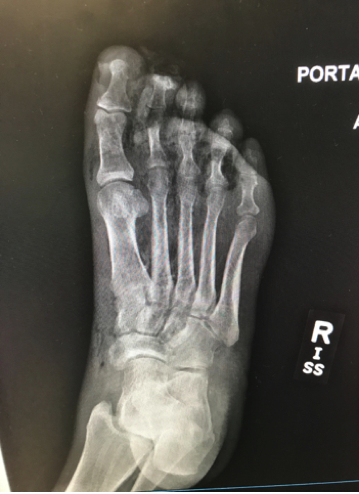
Necrotizing infection of the foot
https://radiopaedia.org/articles/necrotising-fasciitis
Yaghoubian et al. Use of admission serum lactate and sodium levels to predict mortality in necrotizing soft-tissue infections. Archives of surgery. 2007.
Anaya DA and Dellinger EP. Necrotizing soft-tissue infection: diagnosis and management. Clinical infectious diseases. 2007.
Category: Pharmacology & Therapeutics
Posted: 4/27/2017 by Tu Carol Nguyen, DO
Click here to contact Tu Carol Nguyen, DO
Haloperidol has a higher D2 receptor antagonist effect than standard antiemetic treatment agents such as metoclopramide. In addition, newer antipsychotic agents such as Olanzapine have a high affinity at multiple antiemetic sites such as the dopamine and serotinergic receptors.
While formal RCT's are still in the works, multiple sources including palliative care, emergency medicine, and pain journals support their use in refractory emesis.
Consider Haloperidol 3-5 mg IV.
Check an EKG for long QTc prior to use. Consider dose reduction of haloperidol in those with hepatic impairment. Also consider dose reduction in patients taking carbamazepine, phenytoin, phenobarbital, rifampicin, or quinidine due to that pesky CYP3A4 inhibition.
Consider Olanzapine 2-5 mg IV.
Several case reports have shown a higher rate of success with olanzapine for refractory emesis. Olanzapine has similar precautions as those to haloperidol (EKG, hepatic impairment), although it's CYP drug interactions are less common. Additionally, use olanzapine cautiously in hyperglycemic patients as there are several case reports of olanzapine prompting episodes of DKA. Consider frequent blood sugar checks or small doses of insulin in hyperglycemic patients.
Take Home Points:
Consider the antipsychotic agents Haloperidol or Olanzapine for patients with refractory emesis, they may be more effective than traditional antiemetics.
Get an EKG prior to administration to check for QTc prolongation. As the classical and atypical antipsychotic agents are sedating, use caution in conjunction with other sedating medications (such as benzodiazepines).
Category: Visual Diagnosis
Keywords: Pleural effusion; POCUS (PubMed Search)
Posted: 4/17/2017 by Tu Carol Nguyen, DO
Click here to contact Tu Carol Nguyen, DO
A 50 years old male with a history of CHF, presenting to the ED with progressively worsening shortness of breath. POCUS was performed. The picture shows the left lower part of the chest. What is the diagnosis?
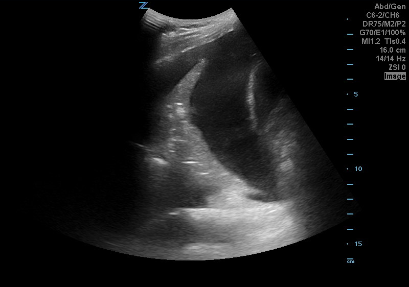
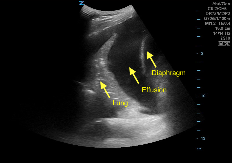
Answer: Pleural effusion
Eibenberger, K. L., Dock, W. I., Ammann, M. E., Dorffner, R., Hörmann, M. F., & Grabenwöger, F. (1994). Quantification of pleural effusions: sonography versus radiography. Radiology, 191(3), 681-684.
Atkinson, P., Milne, J., Loubani, O., & Verheul, G. (2012). The V-line: a sonographic aid for the confirmation of pleural fluid. Critical ultrasound journal, 4(1), 19.
Category: Visual Diagnosis
Posted: 2/13/2017 by Tu Carol Nguyen, DO
Click here to contact Tu Carol Nguyen, DO
56 year-old male with history of hypertension presents with complaints of right scrotal swelling and pain. Denies any urinary symptoms, abdominal pain, nausea/vomiting or change in bowel habits or prior episodes. Temp was 99.0.
A scrotal ultrasound was done and an image of the right testis was seen (below). What's the diagnosis?
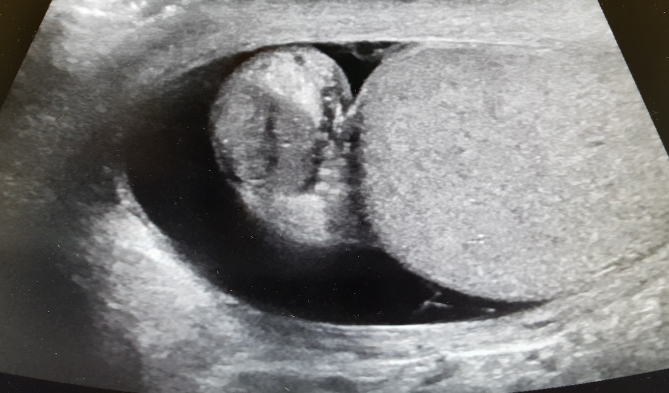
Answer: Right Epididymitis (and Hydrocele)
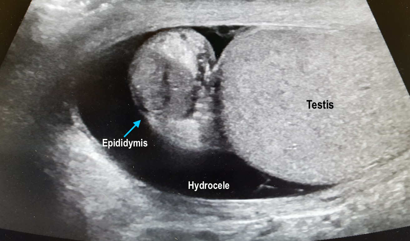
Take Home Points:
Kühn AL, Scortegagna E, Nowitzki KM, Kim YH. Ultrasonography of the scrotum in adults. Ultrasonography. 2016;35(3):180-97.
Centers for Disease Control and Prevention. Sexually Transmitted Diseases Treatment Guidelines, 2015. MMWR Recomm Rep 2015;64(No. RR-3): 1-137.
Category: Visual Diagnosis
Posted: 1/30/2017 by Tu Carol Nguyen, DO
Click here to contact Tu Carol Nguyen, DO
25 year-old female with hx of cerebral palsy with significant developmental delay, s/p G-tube who presented with acute hypoxic respiratory failure, hypotension and a distended, tense abdomen. A CT was done with the scout film below. What's the diagnosis?
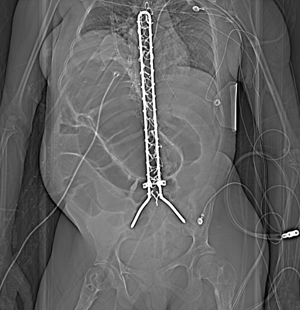
Answer: Cecal Volvulus
Patient was subsequently intubated, had an NG tube placed with over 500cc of fluid returned. Patient had multiorgan failure and received fluids, antibiotics, pressors, blood products and went to the OR, and had a partial bowel resection.
One way to differentiate cecal volvulus from sigmoid volvulus is that sigmoid volvulus generally do not have haustra.
Tonerini M, Pancrazi F, Lorenzi S, Pacciardi F, Ruschi F, et al. (2015) Cecal volvulus: what the radiologist needs to know. Glob Surg, 1: DOI: 10.15761/GOS.1000106.
Category: Visual Diagnosis
Posted: 1/9/2017 by Tu Carol Nguyen, DO
Click here to contact Tu Carol Nguyen, DO
A 60 year-old man with history of atrial fibrillation, CAD presents with left lower leg/foot pain for a few days. His foot is seen below. What's the diagnosis?
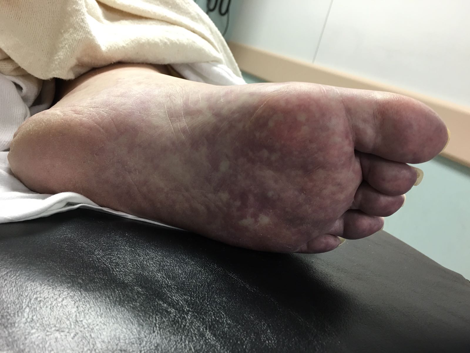
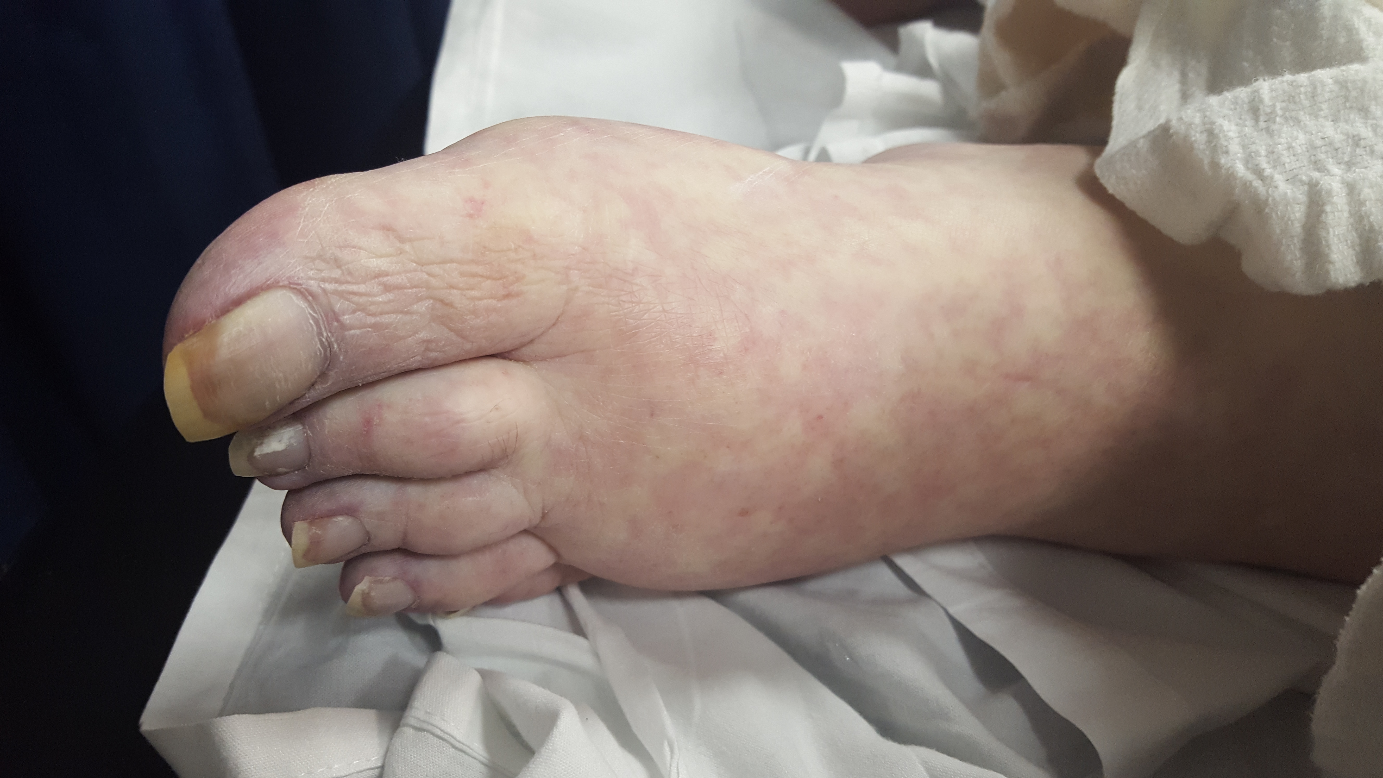
Mottling with concerns for Acute Limb Ischemia
Key Points:
Creager MA, Kaufman JA, Conte MS. Clinical practice. Acute limb ischemia. N Engl J Med. 2012;366(23):2198-206.
http://www.emdocs.net/acute-limb-ischemia-pearls-pitfalls/
Category: Visual Diagnosis
Posted: 12/27/2016 by Tu Carol Nguyen, DO
Click here to contact Tu Carol Nguyen, DO
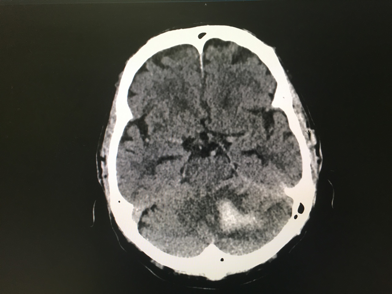
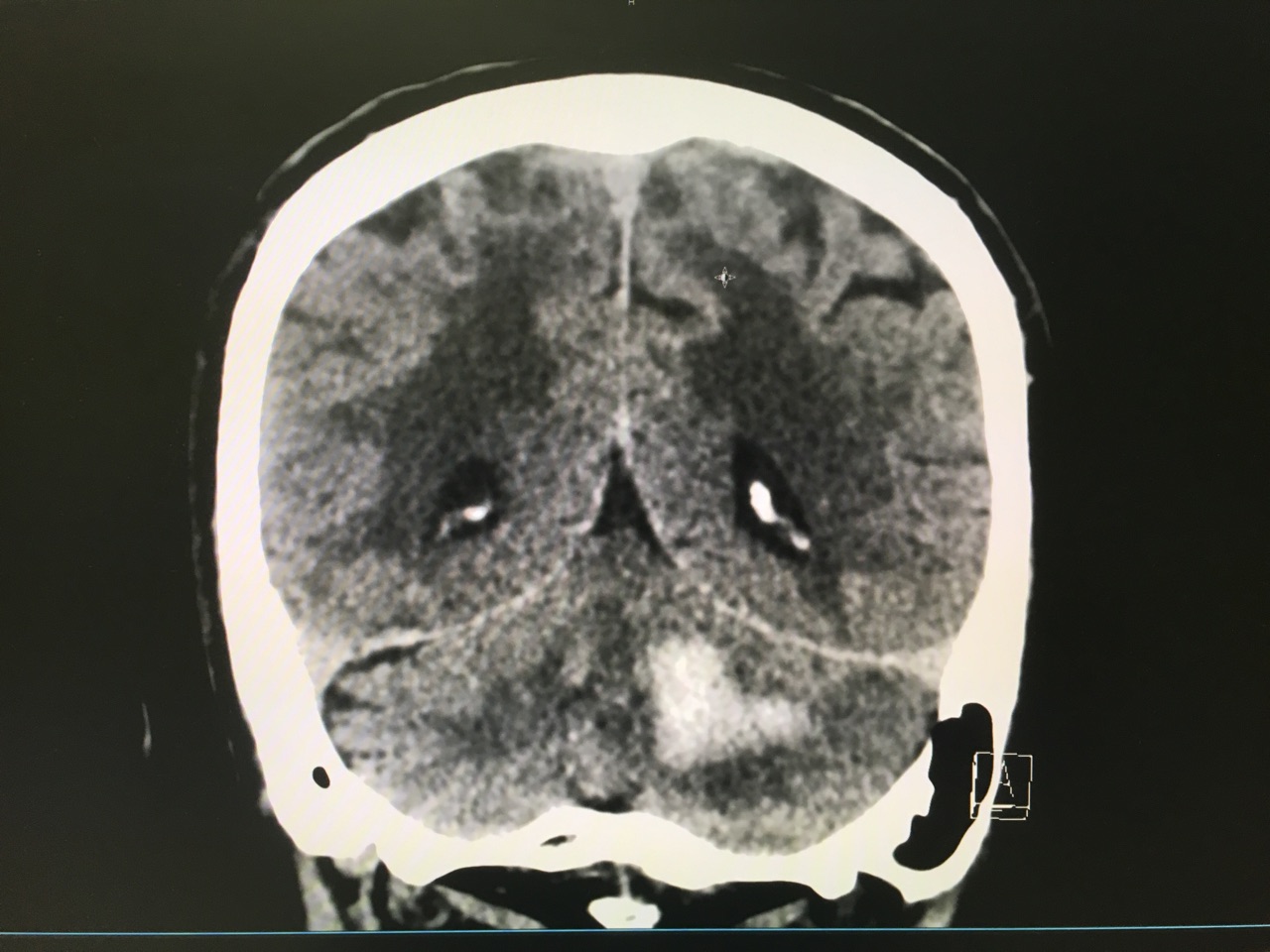
Answer: Large left cerebellar intraparenchymal hemorrhage with surrounding vasogenic edema with mild infratentorial midline shift of ~4mm
Take Home Points:
Hemphill JC, Greenberg SM, Anderson CS, et al. Guidelines for the Management of Spontaneous Intracerebral Hemorrhage: A Guideline for Healthcare Professionals From the American Heart Association/American Stroke Association. Stroke. 2015;46(7):2032-60.
Category: Visual Diagnosis
Posted: 12/5/2016 by Tu Carol Nguyen, DO
Click here to contact Tu Carol Nguyen, DO
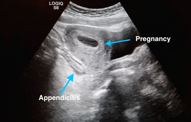
27 year-old G2P1 presents with 3 days of abdominal pain that is mostly suprapubic. Denies any urinary symptoms and vaginal bleeding. Physical examination reveals slight rebound in the right lower quadrant.
An ultrasound revealed the following. What's the diagnosis?
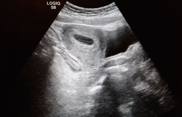
Pregnancy with Appendicitis
Take Home Points:
See previous pearl for how to conduct an ultrasound to evaluate for appendicitis.
Dewhurst C, Beddy P, Pedrosa I. MRI evaluation of acute appendicitis in pregnancy. J Magn Reson Imaging. 2013;37(3):566-75.
Israel GM, Malguria N, Mccarthy S, Copel J, Weinreb J. MRI vs. ultrasound for suspected appendicitis during pregnancy. J Magn Reson Imaging. 2008;28(2):428-33.
Segev L, Segev Y, Rayman S, Nissan A, Sadot E. The diagnostic performance of ultrasound for acute appendicitis in pregnant and young nonpregnant women: A case-control study. Int J Surg. 2016;34:81-85.
Segev L, Segev Y, Rayman S, Nissan A, Sadot E. Acute Appendicitis During Pregnancy: Different from the Nonpregnant State?. World J Surg. 2016.
Theilen LH, Mellnick VM, Longman RE, et al. Utility of magnetic resonance imaging for suspected appendicitis in pregnant women. Am J Obstet Gynecol. 2015;212(3):345.e1-6.
Category: Visual Diagnosis
Posted: 11/7/2016 by Tu Carol Nguyen, DO
Click here to contact Tu Carol Nguyen, DO
8 year-old female with no PMH who presents with concerns for "purple patches" popping up on her arm for 2-3 days. Stated that one appeared and then, the other one appeared 12 hours later. She denied any trauma whatsoever, history of easy bleeding/bruising and did feel safe at home. The rest of the review of systems was negative.
Patient said there was mild pain when the area was touched. The rest of the physical examination was normal.
What's the diagnosis? (Image below)
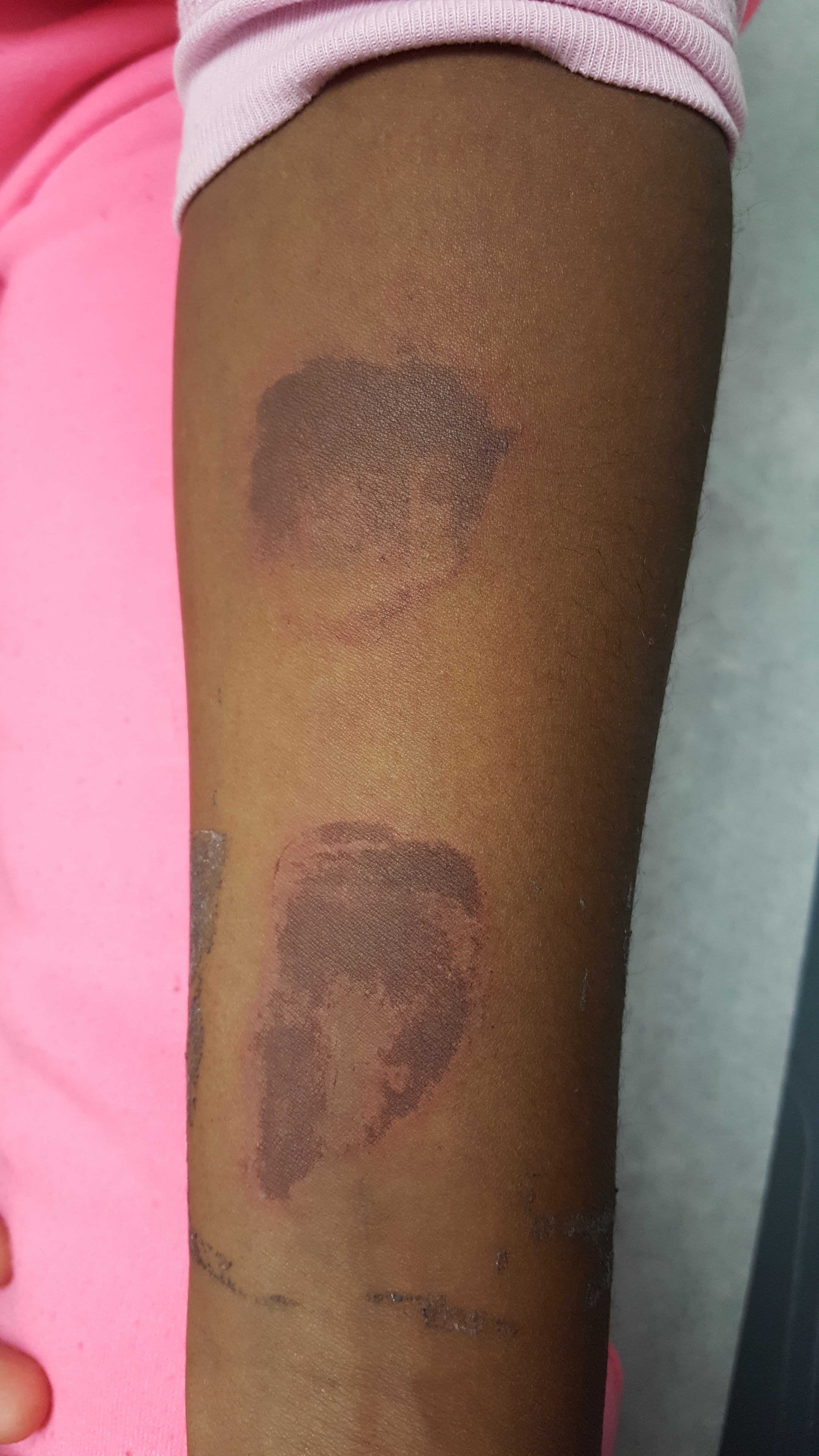
Superficial Thermal Burn
Upon further questioning, patient stated that she had been making s'mores by roasting marshmallows over an electric stove 3 days prior. The burns showed up the subsequent morning.
Take Home Points:
Previous pearls about burns:
Pediatric Burns:
Monseau AJ, Reed ZM, Langley KJ, Onks C. Sunburn, Thermal, and Chemical Injuries to the Skin. Prim Care. 2015;42(4):591-605.
"Pathophysiology of Thermal Injury." Civic Plus. 2007.
Category: Visual Diagnosis
Posted: 10/25/2016 by Tu Carol Nguyen, DO
(Updated: 10/26/2016)
Click here to contact Tu Carol Nguyen, DO
20 year-old female presents with sore throat, right throat fullness, difficulty speaking for 2-3 days. A bedside ultrasound and subsequent CT was obtained as seen below. What's the diagnosis?
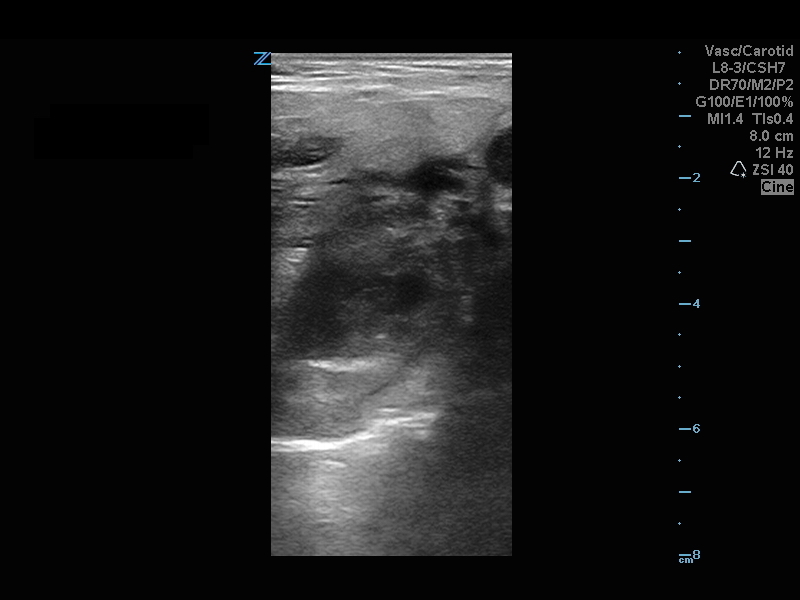
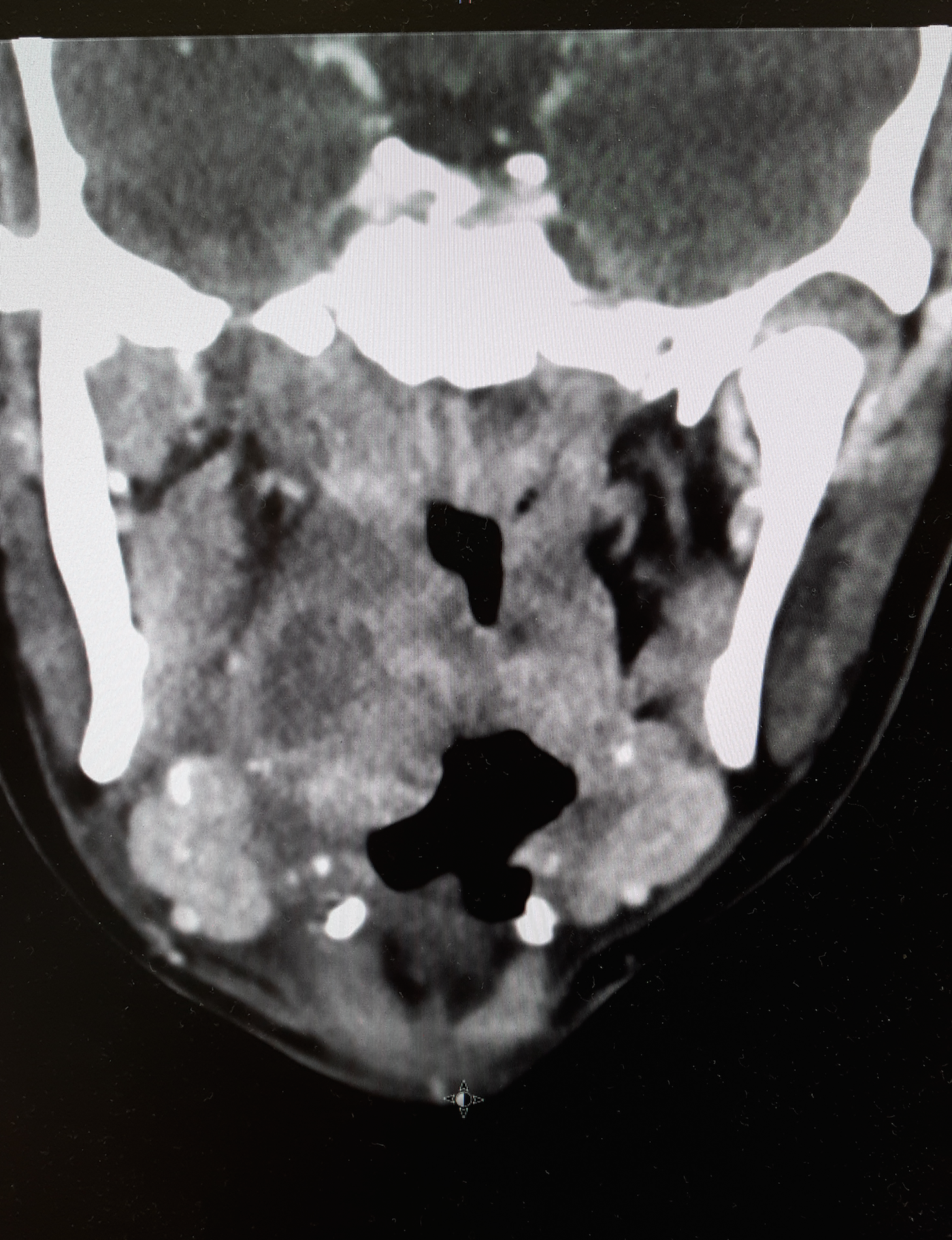
Peritonsillar Abscess
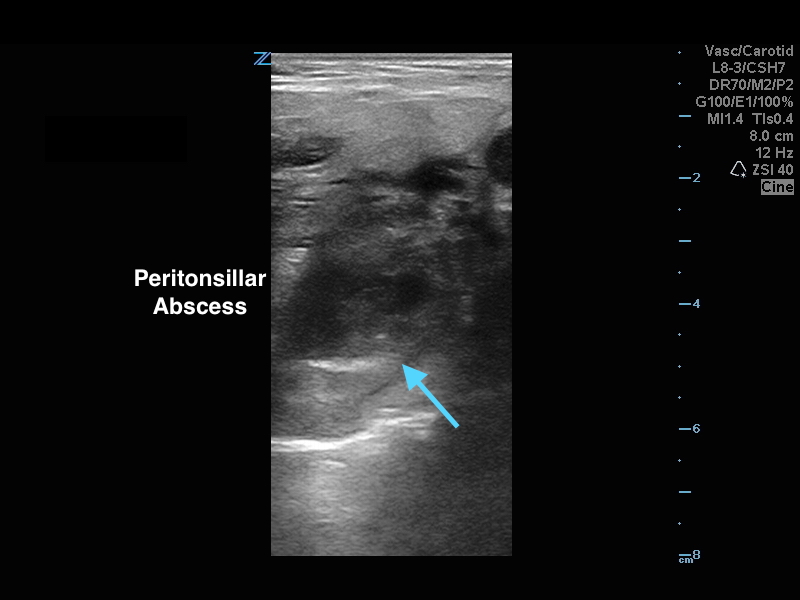
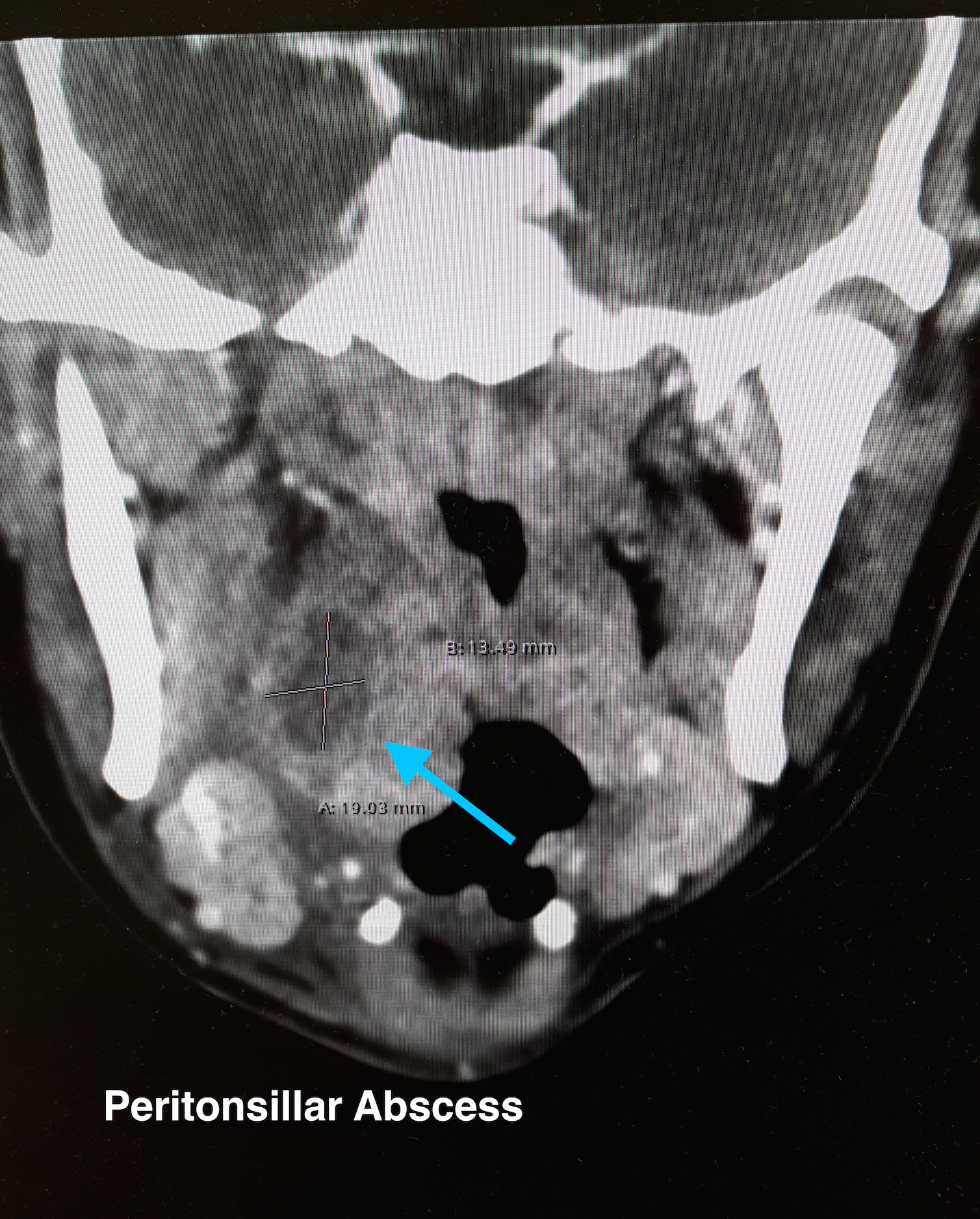
The ultrasound image is a transcutaneous approach with a linear transducer that is placed at the angle of the mandible of the affected side. This is an alternative approach to an intra-oral ultrasound with the endocavitary transducer if the patient has trismus.
Take Home Points:
How to do an intra-oral US-guided needle aspiration of PTA, check out:
http://www.ultrasoundpodcast.com/2012/01/episode-21-full-peritonsillar-abscess-podcast/
For a brief video on how to perform a transcutaneous US for PTA:
https://www.youtube.com/watch?v=JkIYOhKCweI&t=28s
Halm BM, Ng C, Larrabee YC. Diagnosis of a Peritonsillar Abscess by Transcutaneous Point-of-Care Ultrasound in the Pediatric Emergency Department. Pediatr Emerg Care. 2016;32(7):489-92.
Rehrer M, Mantuani D, Nagdev A. Identification of peritonsillar abscess by transcutaneous cervical ultrasound. Am J Emerg Med. 2013;31(1):267.e1-3.
Category: Visual Diagnosis
Posted: 10/10/2016 by Tu Carol Nguyen, DO
Click here to contact Tu Carol Nguyen, DO
57 year-old female with history of bilateral lung transplants presents with fever, and 2 days of a painful, red, bumpy rash over the left labia and left buttock, but also notes a small tender area on the plantar surface of the left foot.
Below is a figure depicting the location of the rash, as well as a photo of her foot.
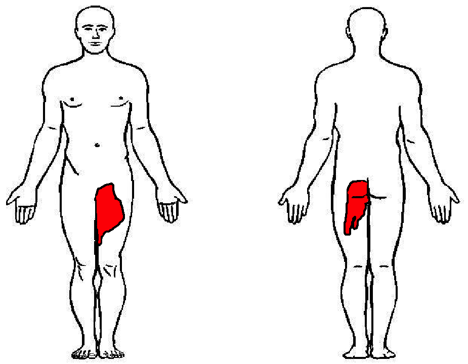
This is Herpes Zoster (Shingles).
Presentation:
Treatment:
Dworkin RH, Johnson RW, Breuer J, et al. Recommendations for the management of herpes zoster. Clin Infect Dis. 2007;44 Suppl 1:S1.
Category: Visual Diagnosis
Posted: 9/14/2016 by Tu Carol Nguyen, DO
(Updated: 9/26/2016)
Click here to contact Tu Carol Nguyen, DO
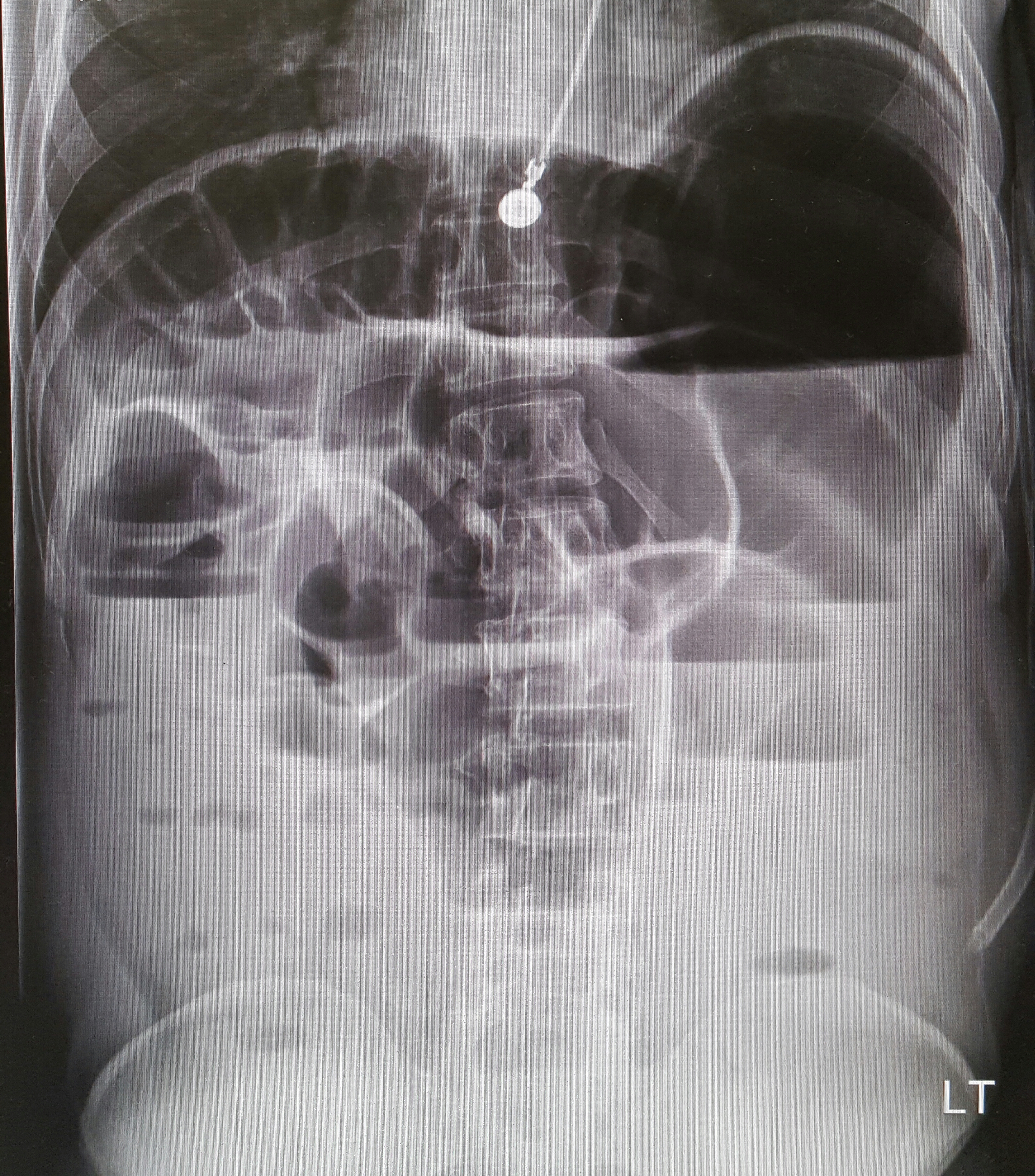
22-year-old male with history of autism, mental retardation who is non-verbal presents with abdominal pain and vomiting for one day. Patient was found clutching his abdomen and moaning. What's the diagnosis?
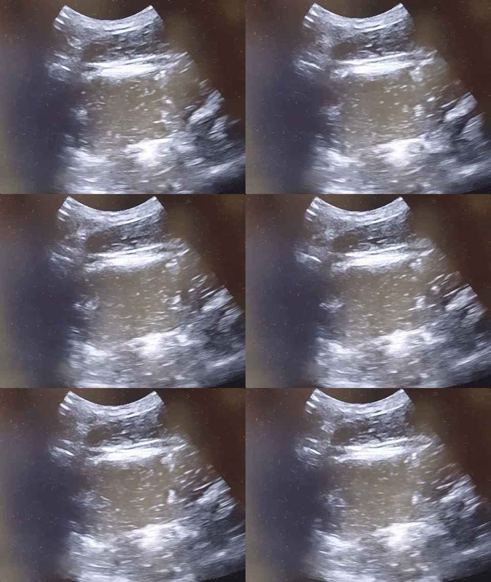
Small Bowel Obstruction
See the corresponding upright abdominal x-ray, showing dilated bowel with air fluid levels.
Kameda T, Taniguchi N. Overview of point-of-care abdominal ultrasound in emergency and critical care. J Intensive Care. 2016 Aug 15;4:53. doi: 10.1186/s40560-016-0175-y. eCollection 2016. Review.
Unl er EE, Yava i O, Ero lu O, Yilmaz C, Akarca FK. Ultrasonography by emergency medicine and radiology residents for the diagnosis of small bowel obstruction. Eur J Emerg Med. 2010 Oct; 17(5):260-4.
Category: Visual Diagnosis
Posted: 9/8/2016 by Tu Carol Nguyen, DO
(Updated: 9/12/2016)
Click here to contact Tu Carol Nguyen, DO
A 25-year-old male was brought in by EMS with a stab wound to the chest. What's the diagnosis?
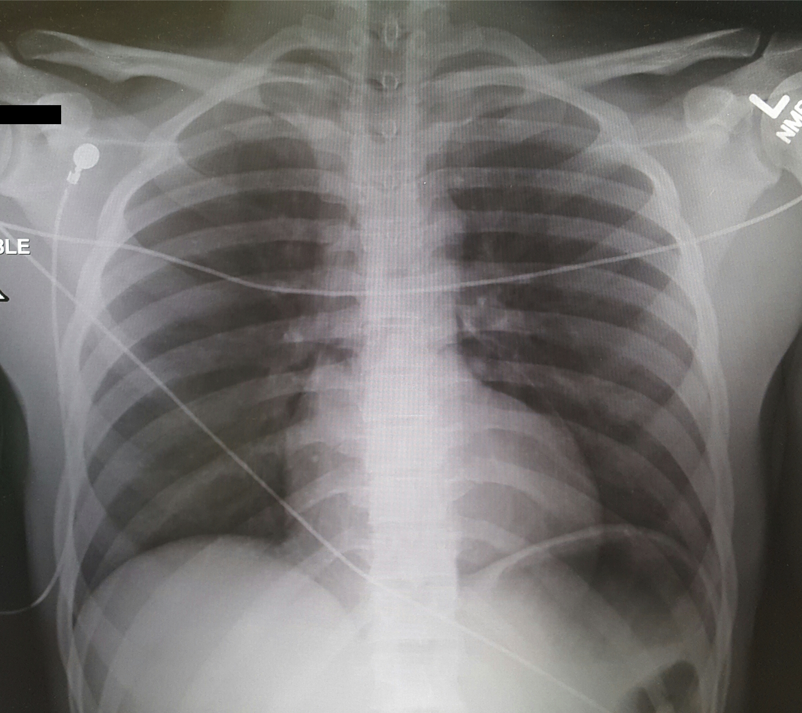
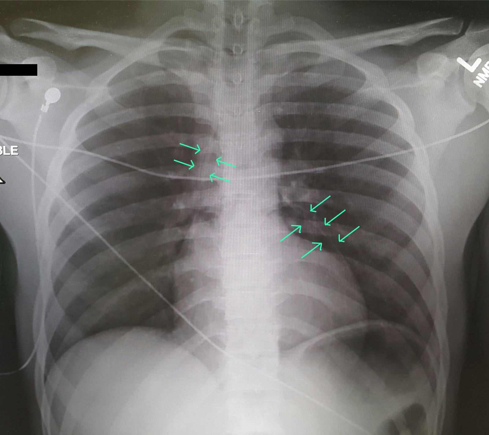
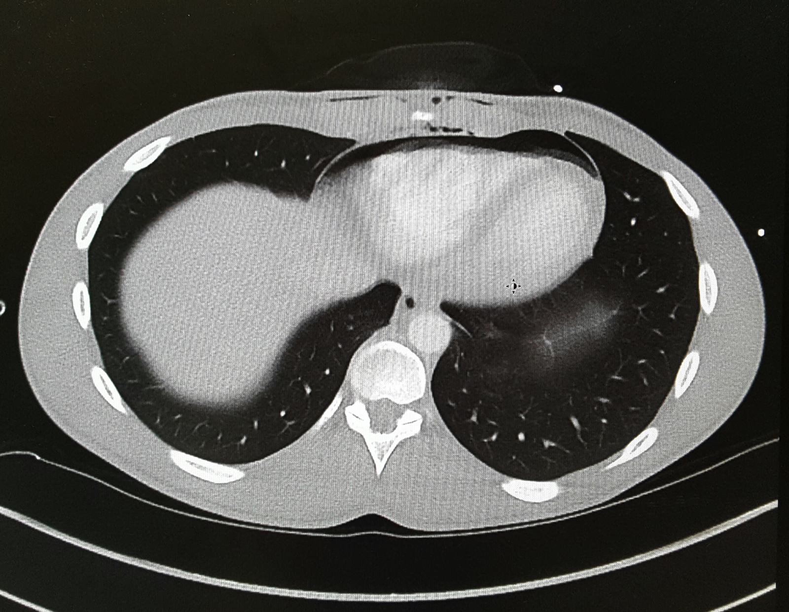
Differential Diagnosis: Bronchial injury, Diaphragm injury, Hemothorax, Tension Pneumothorax, Aortic Transection, Esophageal injury, Pneumomediastinum
Evaluation: Ultrasound (FAST Exam), CXR, CTA in stable patients, ECG, troponin.
Management: Penetrating cardiac trauma require emergent thoracotomy, pericardial window.
Clancy K, Velopulos C, Bilaniuk JW, et al. Screening for blunt cardiac injury: an Eastern Association for the Surgery of Trauma practice management guideline. J Trauma Acute Care Surg. 2012;73(5 Suppl 4):S301-6.
El-menyar A, Al thani H, Zarour A, Latifi R. Understanding traumatic blunt cardiac injury. Ann Card Anaesth. 2012;15(4):287-95.
Tintinalli's 7th Edition. Emergency Medicine Manual. Chapter 164: Cardiothoracic Trauma.
