UMEM Educational Pearls - By Michael Bond

Category: Orthopedics

Title: High Pressure Injection Injuries

Keywords: High Pressure, Injection, Injury (PubMed Search)

Posted: 6/20/2009 by Michael Bond, MD (Updated: 4/23/2024)
Click here to contact Michael Bond, MD

High Pressure Injection Injuries:

  • These injuries initially often have a pretty benign appearance which may result in the injuried person seeking medical treatment late, or the initial medical provider not recognizing the seriousness of the injury.
  • Even when treated promptly and aggressively most patients will end up with an amputation of thier finger or have permanent loss of funciton, strength, sensation, or chronic pain.
  • In a couple of hours, these injuries tend to result in significant swelling that can lead to compartment syndrome. The swelling can be due to the actual disruption of cells from the high pressure, or due to toxic effects of the injected agent.
  • Initial Management should consist of:
    • X-rays: Help to evaluate the extent of the injection.  Radio-opaque solvents will be seen on x-ray, but even radio-lucent solvents may be seen as lucency or air on the x-ray
    • Broad Spectrum antibiotics to prevent infection
    • Corticosteroids to decrease the inflammatory response brought on by the injected agent
    • Tetanus Prophylaxis if needed
    • Emergent hand surgery referral
  • Most if not all patients will require emergent debridement of the affected area.

Show References


Category: ENT

Title: Mandibular Dislocations

Keywords: Mandible, Dislocation, Unified, Hand (PubMed Search)

Posted: 6/13/2009 by Michael Bond, MD (Updated: 4/23/2024)
Click here to contact Michael Bond, MD

Manibular Dislocations:

  • Mandibular dislocations can be extremely difficult to reduce at times.
  • The classic method of reducing a mandible dislocation is for the provider to wrap his thumbs in guaze (to prevent them from being bitten), and while placing his thumbs bilateraly as far posterior on the mandible as possible, he applies downward, and then posterior pressure to reduce the dislocation.
  • Significant muscle spasms can result from the dislocation, requiring procedural sedation, but even with sedation it can be very difficult if not impossible to reduce the mandible.
  • Dr. Cheng's article, referenced below, describes a new technique, where the provider use both of his thumbs to press down on a single side of the mandible posterior until the side reduces.
    • For a bilateral dislocation, the technique would be to reduce one side and then the other.

Some authors also recommend using rolled guaze to hold the patient's mouth shut so that they do not inadvertantly dislocate their jaw a second time if they happen to yawn while awakening from their sedation.

Show References


Category: Orthopedics

Title: Shoulder Dislocations -- Treatment

Keywords: shoulder, dislocation, treatment (PubMed Search)

Posted: 6/7/2009 by Michael Bond, MD (Updated: 4/23/2024)
Click here to contact Michael Bond, MD

Shoulder Dislocations -- Treatment

  • Shoulder dislocations once reduced have typically been treated by placing the arm in a sling and swathe which holds the shoulder in adduction and internal rotation. 
  • However, several studies have now shown that placing the arm in a splint with the shoulder adducted and in 10 degrees external rotation helps to prevent recurrent shoulder dislocation. 
  • Patients should remain in the brace/split for 3 weeks.
  • External rotation is not recommended if there is an associated fracture.
  • Some commerical splints are now available to hold the shoulder in external rotation, however, you can make a small strut with plaster or fiberglass to achieve the same result.

Show References


Category: Orthopedics

Title: Nursemaid Elbow

Keywords: Nursemaid, Radial head, dislocation (PubMed Search)

Posted: 5/30/2009 by Michael Bond, MD
Click here to contact Michael Bond, MD

Nursemaid Elbow:

It is typically taught that the way to reduce a nursemaid's elbow is to hold the elbow at 90 degrees, then firmly supinate and flex the elbow. Place your thumb over the radial head and apply pressure as you supinate.(Taken from Sean Fox's Pearl on 7/20/2007)

However, there is a growing body of evidence that is showing that hyperpronating the forearm actually has a higher success rate on first attempt, is easier to perform, and is associated with less pain then supinating the forearm.  The overall reducation rates where similar for both methods.

The hyperpronation method consists of hyperpronating the forearm and then flexing the elbow.  Since the child tends to already hold their arm in partial pronation, the hyperpronation technique tends to need less force and has been associated with less pain.


Show References


Category: Orthopedics

Title: Elbow Dislocations

Keywords: Elbow Dislocation (PubMed Search)

Posted: 5/23/2009 by Michael Bond, MD (Updated: 4/23/2024)
Click here to contact Michael Bond, MD

Elbow Dislocation

  • The elbow is the second most commonly dislocated joint after the shoulder in adults. 
  • It is the most commonly dislocated joint in children.
  • 90% of all elbow dislocation are posterior.  A considerable amount of force is required to dislocate the elbow so be highly suspicous for associated fractures of the radial head, or coronoid process of the ulna. 
  • The combination of a radial head fracture, coronoid process fracture and elbow dislocation is known as the terrible elbow.
  • Anterior elbow dislocations can be associated with injuries to the brachial artery, median and ulnar nerves. 

Quick clinical clues that the elbow is dislocated:

  • Posterior dislocation typically will have a prominent olecranon process, the arm is flexed at the elbow, and the forearm will appear shortened.
  • Anterior dislocation typically present with the arm in extension and the forearm will appear elongated.


Category: Orthopedics

Title: Trimallelor Fracture

Keywords: Trimallelor Fracture (PubMed Search)

Posted: 5/16/2009 by Michael Bond, MD (Updated: 4/23/2024)
Click here to contact Michael Bond, MD

Trimallelor Fractures:

Bimallelor fracture involve both the medial mallelous of the tibia and the distal fibula.  The third malleloi is the posterior tip of the articular surface of the tibia. Can result in instability in the posterior and lateral directions along with external rotation.

Some indications for Open Reduction Internal Fixation when the posterior mallelous is fractured are:

  • > 25% of the posterior articular surface being involved.
  • Fractures that allow posterior subluxation of the talus
  • Fractures that are displaced more than 2 mm
  • Fractures that can not be reduced satisfactorily.


Category: Orthopedics

Title: Knee Dislocation

Posted: 5/9/2009 by Michael Bond, MD (Updated: 4/23/2024)
Click here to contact Michael Bond, MD

Knee Dislocations:

Are relatively rare injuries, but can result in loss of the limb if missed.  Patients will sometimes say they dislocated their knee when they actually mean their patella, so a good history where they describe what their knee looked like, and what they were doing at the time will help differentiated the two.

Some signs that you are dealing with a spontanously reduced knee dislocation are:

  • Varus or valgus instability in full extension of the knee is suggestive of a grossly unstable knee
  • Pain out of proportion to injury
  • Absent or decreased pulse

The loss of limb is due to unrecognized injury to the popiteal artery which as be estimated to occur 7-45% of the time. 

  • Normal pulses and a normal capillary refill does NOT rule out as significant vascular injury. 
  • Arteriograms are no longer mandatory in all cases, but it is generally recommended that you perform an ankle-brachial index and get a vascular duplex scan of the popiteal artery to exclude dissections, tears, aneurysms and psuedo-anuerysms that can all occur as a result of the dislocation.

If you would like to see some videos of knee injuries in the making follow this link www.csmfoundation.org/Educational_Lower_Extremity.html


Category: Orthopedics

Title: Distal Radius Fractures

Keywords: radius, fracture, colles, smith, barton, chauffer (PubMed Search)

Posted: 5/2/2009 by Michael Bond, MD (Updated: 4/23/2024)
Click here to contact Michael Bond, MD

Distal Radius Fractures

  • The radius is the most commonly fracutred bone of the arm.
     
  • The Colles fracture is a fracture of the distal radius that is angulated dorsally [The distal fragment is angulated towards the back of the hand.]
     
  • The Smith fracture is similar but the distal fracture is angulated volarly [towards the palm of the hand]
     
  • Other less commonly named fractures are the:
    • Barton's - an intraarticular fracture fo the distal radius with dislocation of the radiocarpal joint.  Typically occrus as a fall on the extended and pronated wrist.
       
    • Chauffeur's fracutre - a fracture of the radial styloid process.  Typically caused by compression of the scaphoid against the styloid.  Also known as a hutchinson fracture.


Category: Orthopedics

Title: Phalanx Fractures

Keywords: Phalanx, fracture, treatment (PubMed Search)

Posted: 4/25/2009 by Michael Bond, MD (Updated: 6/27/2009)
Click here to contact Michael Bond, MD

  • Fractures of the phalanx are common, and fractures of the proximal phalanx can lead to significant disability if not treated appropriately.
  • Be sure to check for malrotation, which is a common problem.  Check for this by examing for the normal cascade in finger flexion with the tips of the fingers pointing toward the proximal portion of the scaphoid
  • Acceptable Reduction:
    • No rotational deformity can be accepted
    • No more than 10 deg of angulation should be accepted in any plane
    • Malreduction will cause loss of equilibrium between flexor and extensor tendons.
  • Place the splint on the dorsum side of the finger so that the patient can still have sensation of the tip of their finger tip.
  • Patients requiring prompt referral to a hand surgeon are those with:
    • Intraarticular fractures
    • Malrotation
    • Unacceptable reductions
    • Unstable fractures


Category: Airway Management

Title: Le Fort Fractures

Keywords: Le Fort, fracture, facial (PubMed Search)

Posted: 4/19/2009 by Michael Bond, MD (Updated: 8/28/2014)
Click here to contact Michael Bond, MD

The French Surgeon Rene Le Fort first described these facial fracture patterns.   Reportedly he made the observations after dropping numerous skulls from the wall of a castle.  This might be why we don't see pure Le Fort fractures in our patients most of the time as they are not likely to be falling off castle falls head first.

The classic fracture patterns are:

  1. Le Fort I fractures extends from the nasal septum to the lateral pyriform rims, travels horizontally above the teeth apices, crosses below the zygomaticomaxillary junction, and traverses the pterygomaxillary junction to interrupt the pterygoid plates.
  2. Le Fort II fracture has a pyramidal shape and extends from the nasal bridge at or below the nasofrontal suture through the frontal processes of the maxilla, inferolaterally through the lacrimal bones and inferior orbital floor and rim through or near the inferior orbital foramen, and inferiorly through the anterior wall of the maxillary sinus; it then travels under the zygoma, across the pterygomaxillary fissure, and through the pterygoid plates.
  3. Le Fort III fractures (transverse) are otherwise known as craniofacial dissociation and involve the zygomatic arch.  These fractures start at the nasofrontal and frontomaxillary sutures and extend posteriorly along the medial wall of the orbit through the nasolacrimal groove and ethmoid bones. The fracture continues along the floor of the orbit along the inferior orbital fissure and continues superolaterally through the lateral orbital wall, through the zygomaticofrontal junction and the zygomatic arch.


http://radiographics.rsnajnls.org/cgi/content-nw/full/26/3/783/F15


Category: Misc

Title: G6PD Deficiency

Keywords: G6PD, Deficiency (PubMed Search)

Posted: 4/11/2009 by Michael Bond, MD (Updated: 4/23/2024)
Click here to contact Michael Bond, MD

Glucose-6-Phosphate Dehydrogenase Deficiency

  • G6PD Deficiency is a genetic disorder which can cause hemolytic anemia when people with the disorder come into contact with drugs, food and other substances which cause oxidative stress.
  • It is the most common genetic enzyme deficiency.
  • G6PD is an inherited disorder with over 400 different known variants.
  • Oxidative stress can cause the premature distruction of RBC's due to the lack of the enzyme reduced glutathione which G6PD helps produce.
  • Drugs that are at high risk for causing hemolytic anemia in those with G6PD deficiency are:
    • NSAIDS (Asprin, Tylenol, Ibuprophen)
    • Quinolones
    • Sulfa drugs
    • Drugs metabolized known to cause blood or liver related problems or hemolysis
    • Primaquine
    • Nitrofurantoin
    • Glyburide
    • Dapsone

Also make sure that you are not G6PD deficiency if you are eating with Hannibal Lecter as Fava beans and other legumes can also cause an episode of hemolysis.

A good reference for G6PD deficiency is http://g6pddeficiency.org/index.php


Category: Orthopedics

Title: Radial Head Fractures

Keywords: Radial, Head, Fracture (PubMed Search)

Posted: 4/3/2009 by Michael Bond, MD (Emailed: 4/4/2009) (Updated: 4/23/2024)
Click here to contact Michael Bond, MD

Radial Head Fractures:

Radial head fractures are more common in adults, where radial neck fractures are more common in children.  Remember to look for fat pads to help make the diagnosis if it is not obvious on plain films.  On plain films, a line drawn down the middle of the radial head should always line up with the capitellum of the humerus.  If this does not occur the radial head is dislocated and/or fracture.

Orthopaedics use the Mason classification to help guide treatment, and break down fractures into 3 different types.

  • Type I - is undisplaced, generally treated nonoperatively. 
    • Early mobilization prevents chronic elbow stiffness.
  • Type II - a single fragment is displaced.
    • May be treated nonoperatively if the displacement is minimal.
    • The rule of threes is used. Nonsurgical treatment can be considered if the fracture involves less than one third of the articular surface, less than 30° of angulation, and if displacement is less than 3 mm
  • Type III  - is comminuted.
    • Usually require operative intervention.


Category: Orthopedics

Title: Hamate Fractures

Keywords: Hamate, Fracture, (PubMed Search)

Posted: 3/28/2009 by Michael Bond, MD (Updated: 4/23/2024)
Click here to contact Michael Bond, MD

Hamate Fractures:

  • Typically the result of a direct blow, and the hook of the hamate is commonly fractured in batters or golfers.
  • Like the scaphoid, the hook is at risk for avascular necrosis and non-union of the hook.
  • Fractures of the body are more common than fracture of the hook of the hamate
  • On exam you will typically find:
    • Increased pain with axial loading of ring (4th) and little finger (5th) metacarpals
    • Most patients complain of pain and tenderness on ulnar side of palm or on the dorsoulnar aspect of the wrist.
    • Pain also aggravated by grasping items.
  • Diagnosis
    • Fracture often missed on routine AP & lateral films
    • Most fractures can be diagnosed by plain films if you as for the "Carpal tunnel view"
    • CT scan can also be used to see the fracture
  • Treatment
    • Good Immobilization will often prevent avascular necrosis and allow early healing
      • Volar splint or short arm cast are usually adequate.
    • Excision of the hook of the hamate provides similar results as an ORIF in those that have non-union or displaced fractures.
    • Refer to orthopedics

Show References


Category: Orthopedics

Title: Lunate Dislocation

Keywords: Lunate, Dislocation, Perilunate (PubMed Search)

Posted: 3/20/2009 by Michael Bond, MD (Emailed: 3/21/2009) (Updated: 4/23/2024)
Click here to contact Michael Bond, MD

Lunate Dislocation and perilunate dislocation are broken down into 4 stages that relates to the progressive disruption of the carpal ligaments due to hyperextension and ulnar deviation of the wrist:

  • Stage 1: Scapholunate Dislocation
    • Has the characteristic sign of widening of the scapholunate joint on the PA view known as the Terry Thomas Sign as it resembles the gap between his teeth
    • Gap between scaphoid and lunate should be less than 2 mm
  • Stage II: Perilunate dislocation
    • Best seen on lateral view of the wrist
    • Associated with scaphoid fractures
    • Lunate stays in its normal position with the capitate dislocation posterior when you use the distal radius as your reference point
  • Stage III: Perilunate dislocation
    • Also includes dislocation or fracture of the triguetrum
    • Triquetrial and scaphoid malrotation
    • In lateral view, all other carpal bones are dislocated posterior with respect to lunate
  • Stage IV:  Lunate dislocation
    • On PA view you will see a triangular view of the lunate on the PA view that looks like a "piece of pie". 
    • On the lateral view of the wrist the lunate will look like a tea cup tipped in the volar direction AKA the "spilled teacup sign"
    • Associated with a scaphoid fracture
       

For a good indepth review of lunate and perilunate injuries please read the article by Andy Perron with this attached link.... doi:10.1053/ajem.2001.21306   

If you are interested in seeing some xray examples please visit LearningRadiology.com


Show References


Category: Orthopedics

Title: Fractures and Child Abuse

Keywords: Child Abuse, Fracture (PubMed Search)

Posted: 3/15/2009 by Michael Bond, MD (Updated: 4/23/2024)
Click here to contact Michael Bond, MD

A lot of what is taught about fracture patterns in abused children has been extrapolated from post-mortem studies which is a different population then what you will see in the Emergency Department. The study referenced did a metanalysis of all the literature in an attempt to determine what fractures suggest abuse and looked at all comers that had fractures.  Some of the patterns they were able to extrapolate are:


  • Fractures from abuse predominately occurred in infants and toddlers
    • In children less than 12 one study showed that 80% of all fractures from abuse occurred in children less than 18 months old.
    • In children over 5 years old 85% of fractures are not caused by abuse
  • In children under 3 years old, skull fractures were by far the most common fracture type in both abused and non-abused children.
    • However, the presense of a skull fracture only has a 1:3 chance of being from abuse.
    • Skull fractures location and type are similar between abuse and non-abuse, though multiple fractures and fractures that cross suture lines are more highly associated with abuse.
  • There is a strong relationship between multiple fractures and abuse
    • 74% of abused children had two or more fractures compared to 16% of non-abused
  • In the absence of a confirmed traumatic case, rib fractures have the highest probability (71%) of being caused by abuse.
  • Humeral fractures have a 1:2 chance of being the result of abuse.
  • Femur fracture like skull fractures have a 1:3 chance of being the result of abuse.


Show References


Category: Orthopedics

Title: Galeazzi Fracture

Keywords: Galeazzi, Fracture (PubMed Search)

Posted: 3/7/2009 by Michael Bond, MD (Updated: 4/23/2024)
Click here to contact Michael Bond, MD

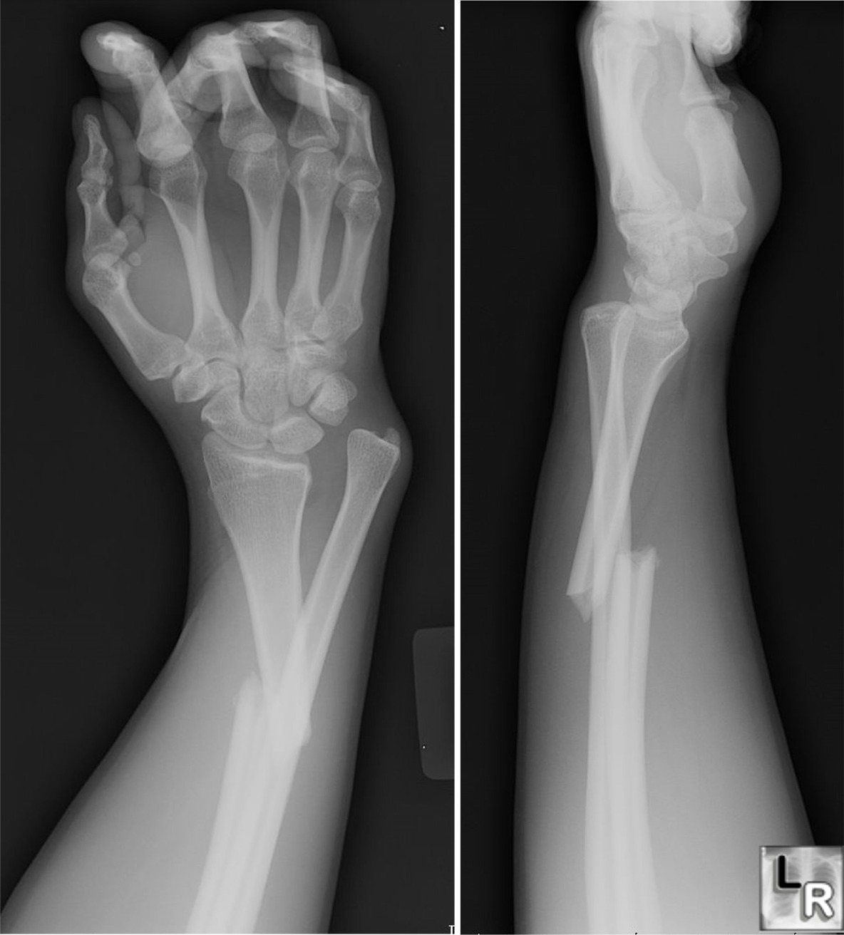
The Galeazzi Fracture:

  • It is a fracture of the distal to middle third of the radial shaft with dislocation of the Distal Radio-Ulnar Joint. 
  • Typical mechanism of injury is a fall onto a outstretched hyperpronated forearm.
  • Estimated to represent 7% of adult forearm fractures.
  • This fracture requires surgical repair (Open reduction and internal fixation) in order to prevent presistant or recurrent dislocation of the distal ulnar which typically occurs with closed reduction techniques.
  • Associated with injury to the Anterior interosseous nerve which is a purely motor branch of the median nerve.  Injury results in paralys of the flexor pollicis longus (FPL) and flexor digitorum profundus (FDP) to the index finger, resulting in loss of the pinch mechanism between the thumb and index finger.

To see a photo of a Galeazzi fracture please visit the Learning Radiology Website by clicking on the following link:

http://www.learningradiology.com/caseofweek/caseoftheweekpix2/cow157lg.jpg


Category: Orthopedics

Title: The Ottawa Rules

Keywords: Ottawa, Ankle, Knee, Foot (PubMed Search)

Posted: 2/28/2009 by Michael Bond, MD (Updated: 4/23/2024)
Click here to contact Michael Bond, MD

Most people are familiar with the Ottawa Ankle Rules, but there are also Ottawa Knee and Foot rules.  The Ottawa rules help to limit the number of x-rays you may need in patients that present with ankle, foot or knee pain after an injury.

The Ottawa Ankle Rule

An ankle x-ray is only needed if there pain in the mallelolar area and any of the following:

  • Bone tenderness at the posterior tip of the base of the lateral mallelous
  • Bone tenderness at the posterior tip of the base of the medial mallelous
  • Inability to weight bear immediately and in the Emergency Department

The Ottawa Foot Rule

A foot x-ray is only needed if there is pain in the midfoot and any of the following:

  • Bone tenderness at the base of the 5th metatarsal
  • Bone tenderness over the navicular
  • Inability to weight bear immediately and in the Emergency Department

The Ottawa Knee Rule

A knee x-ray is only needed for knee injury patients when they have any of the following:

  • Age 55 or over
  • Isolated tenderness of the patella (no bone tenderness of the knee other than the patella)
  • Tenderness at the head of the fibula
  • Inability to flex to 90 degrees
  • Inability to weight bear both immediately and in the Emergency Department (4 steps - unable to transfer weight twice onto each lower limb regardless of limping).


Category: Vascular

Title: Bleeding AV Fistulas

Keywords: AV fistulas, bleeding (PubMed Search)

Posted: 2/21/2009 by Michael Bond, MD (Updated: 4/23/2024)
Click here to contact Michael Bond, MD

Bleeding AV Fistulas

It is not an uncommon complaint for dialysis patients to present with bleeding from their fistula.  They can lose a large amount of blood in a short period of time if not treated promptly, and if treated too agressive their fistula can clot off. Some tips on how to control the bleeding.

Most of the bleeding occurs at the site that the needle puntured the fistula. If it is due to an ulcer eroding into the fistula these tips may not be effective.

  • The easiest and safest way to control the bleeding is with simple diret pressure directly over the site of bleeding with a single finger. No guaze.  [Gown up and wear goggles or eye protection]. The use of a big wad of guaze or a pressure dressing tends to just hide the continued bleeding or result in the clotting off of the fistula.
  • Injecting lidocaine with epinephrine at the site can also help and helps set you up for the next step,
  • A figure eight stitch at the puncture site can help close the puncture wound.
  • Of course you should call your vascular surgeon if you are having trouble controlling the bleeding, want close follow up or finger is going numb from holding pressure.

I typically check a CBC and coags.  Once the bleeding is controlled observe the patient for awhile [typically the hour to hour and half to get the labs back] and then road test them with a walk around the Emergency Department to ensure it does not start bleeding again.


Category: Orthopedics

Title: Ankle Sprains

Keywords: Ankle Sprain, Treatment (PubMed Search)

Posted: 2/14/2009 by Michael Bond, MD (Updated: 4/23/2024)
Click here to contact Michael Bond, MD

Ankle sprains are typically treated with a short period of immbolization and then functional exercises are prescribed to rehabilitate the ankle.  A study published in the Lancet this week might just change that.  Lamb et al looked at 584 people with severe ankle sprains (unable to weight bear 3 days out from injury) that were randomized to be treated with a 10 day below knee cast, Aircast, Bledshoe Shoe or Tubular Compression dressing (similar to Ace Wrap).  Those that were treated with the Cast and Aircast had quicker return to function and less disability at 3 months.  There was no increased risk of DVTs in the cast group.

A commentary in the same issue points out that severe ankle sprains are associated with:

  • lower levels of physical activity levels
  • recurrent ankle sprains are often reported for months and years after initial injury.
  • About 30% of patients with an initial ankle sprain develop chronic ankle instability, or repetitive giving way of the ankle during functional activities.
  • There is also emergent evidence to link severe and repetitive ankle sprains to increased risk of ankle osteoarthritis.

Based on this article I think it is prudent to treat all patients with severe Ankle Sprains with a prolonged period of forced immobilzation (Posterior Splint, Short Leg Cast or Aircast).  I would also recommend the Aircast be used to prevent recurrent sprains especially if the patient is involved in sports that require jumping (Basketball, Volleyball) where the risk of reinjury is higher.

Show References


Category: Trauma

Title: Lidocaine with Epinephrine and it use on Fingers and Toes

Keywords: Epinephrine, Lidocaine, Fingers, (PubMed Search)

Posted: 2/7/2009 by Michael Bond, MD (Updated: 4/23/2024)
Click here to contact Michael Bond, MD

Lidocaine with Epinephrine and it use on Fingers and Toes

It has been taught for a long time that Lidocaine with Epinephrine should not be used on fingers, toes, ears and nose [There has to be a kid's song in there somewhere] due to the risk of vasoconstricition/vasospasm and possible digitial infarcation.

The short story is that this practice is not supported by the literature, and there are now numerous publications that have shown that lidocaine with epinephrine is safe for use on the finger tips.  It turns out the the original case reports were submitted with procaine and epinephrine and not lidocaine with epinephrine.  Most of the cases of digital infarction where with straight procaine that is now thought to have been contaiminated or too acidic pH close to 1 when injected.

The effects of epinephrine last approximately 6 hours. This time is well within the accepted limit of ischemia for fingers that has been established in digitial replanation.

So why use Lidocaine with Epinephrine:

  1. Provides a longer period of anesthesia
  2. Decreases bleeding which:
    1. Improves visualization of tendons and underlying structures
    2. Makes repairs easier
    3. Decreases need for a torniquet


Show References