Small Bowel Obstruction
Aortic Dissection
Ultrasound has a great specificity for aortic dissection. Remember to take a look at your aorta on all cardiac views.
Let’s give a shout out to Nikki Cali for diagnosing aortic dissection in a patient with a recent PE. Can you find the dissection flap in this image?
Peritonsillar Abscess


Appendicitis
Ultrasound has a reported high specificity (97.9) for acute appendicitis in moderate to high pre-test probability of patients.
Let’s give a shout out to Reed Macy, who diagnosed appendicitis in a male with vomiting and abdominal pain!
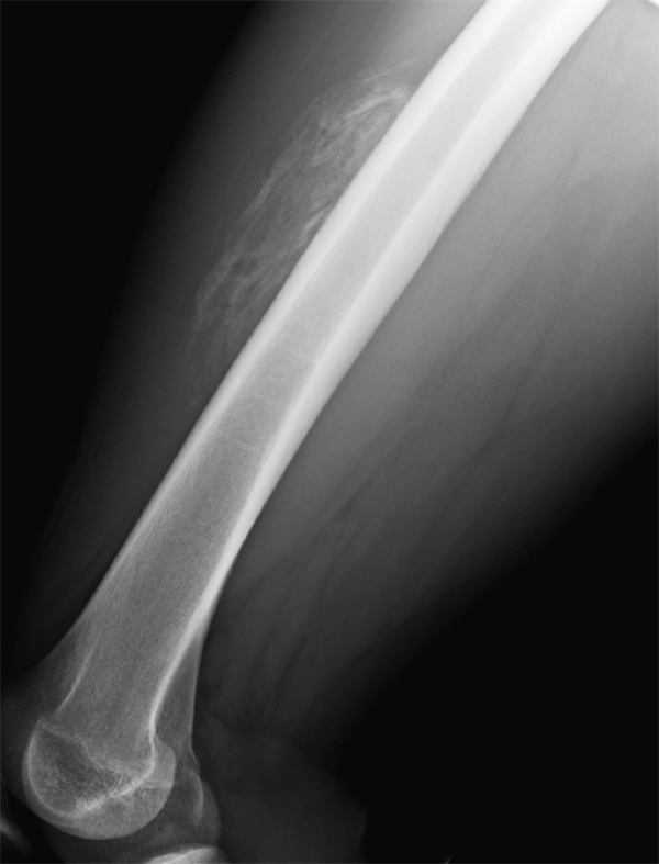
23 y/o otherwise healthy Male presents for approx. 3 month history of Right leg mass. It is painful with activity (deep and sharp) but not enlarging. Patient remembers a fall from a bicycle 6 months ago, with negative imaging for fracture.
What is the diagnosis?
https://plinthsandplatforms.files.wordpress.com/2016/06/screen-shot-2016-06-20-at-10-58-18-am.png
https://radsource.us/wp-content/uploads/2019/02/1E.jpg
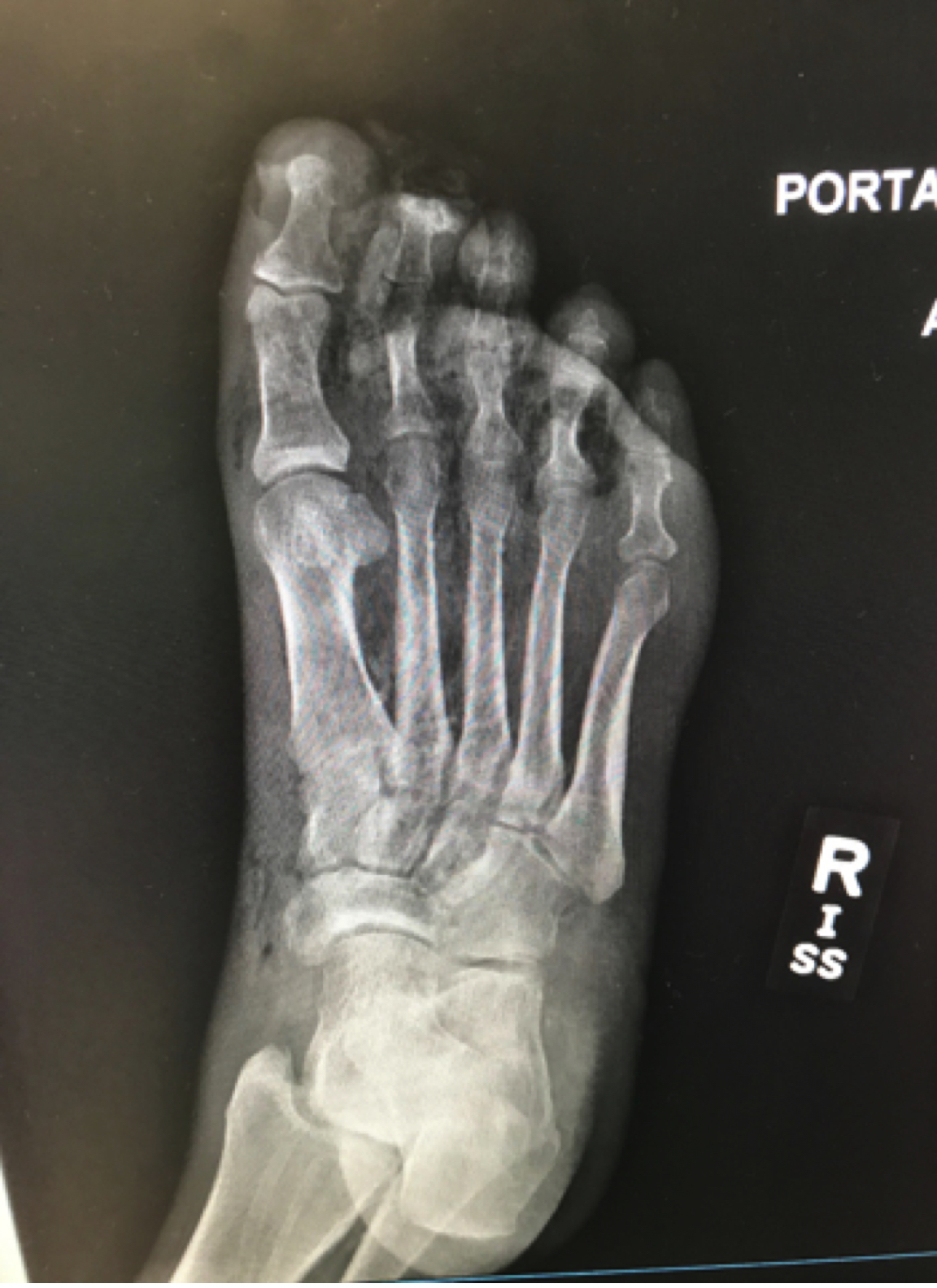
A ~55 year-old female with a history of ESRD and diabetes who presented to the ED with progressively worsening foot odor. An x-ray was performed. The picture below shows the right foot.
What is the diagnosis?
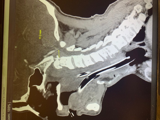
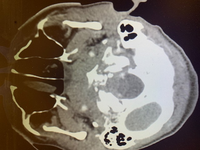
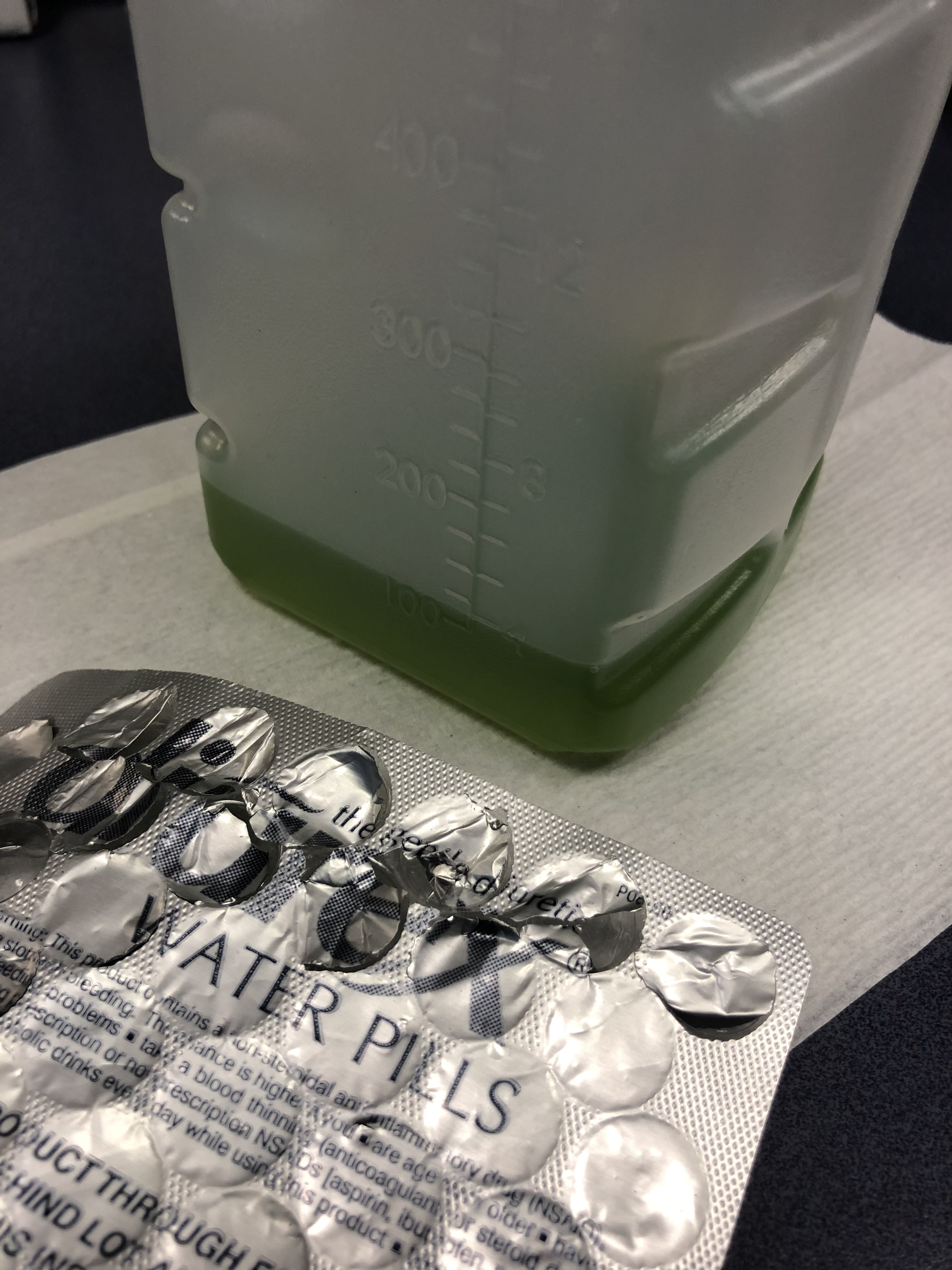
75 y/o M is brought in by EMS after he fell off the light rail and hit his head. In the ED he is A&Ox3, and is asking for a urinal. Two minutes later the tech comes running to show you the following:
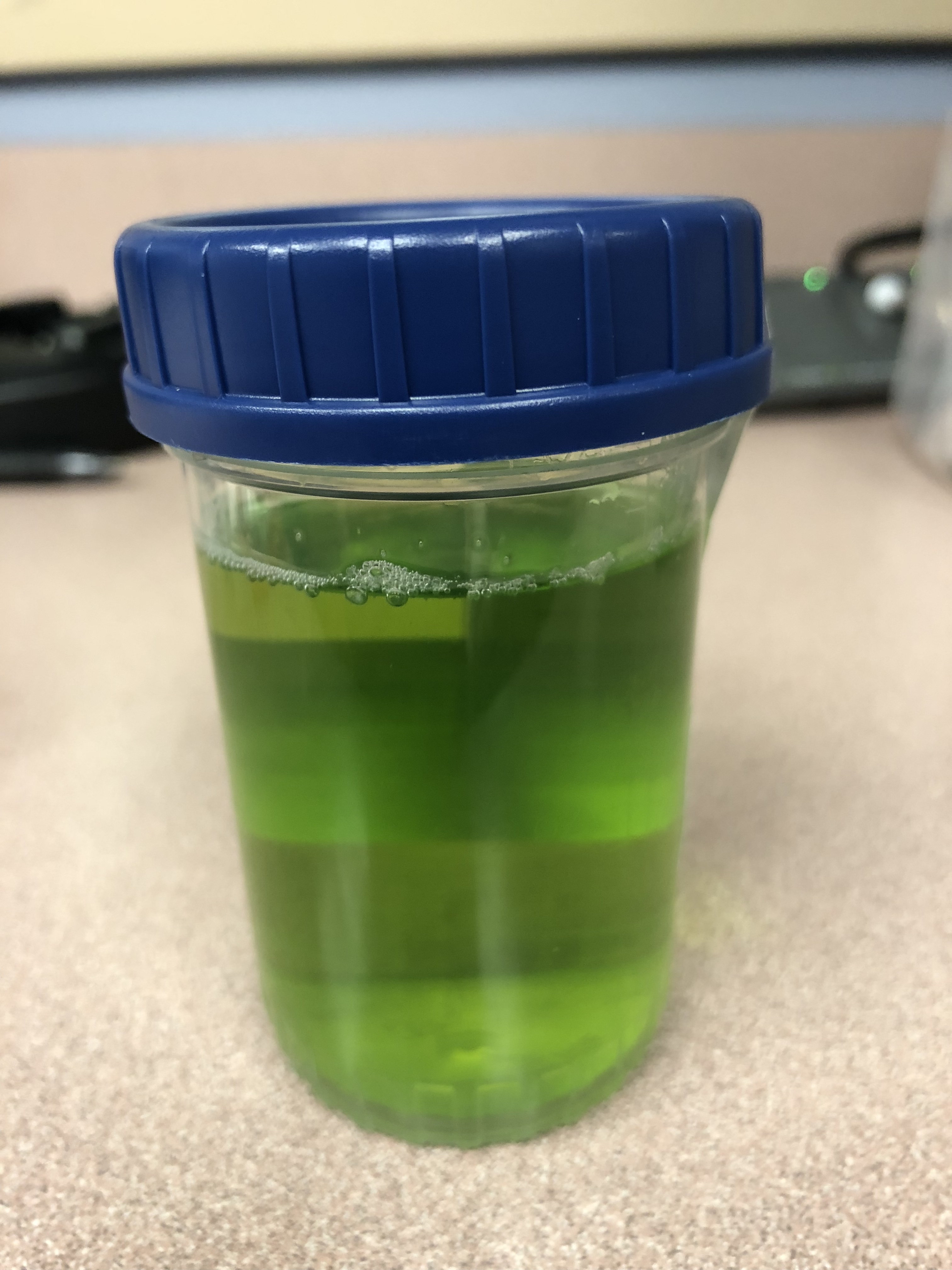
What is the cause of this patients Jolly Rancher Green Apple looking urine sample?
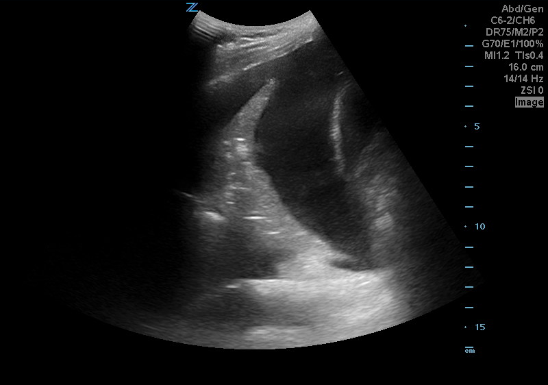
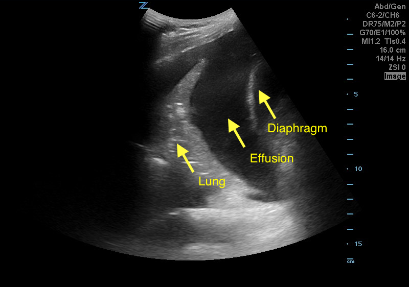
A 50 years old male with a history of CHF, presenting to the ED with progressively worsening shortness of breath. POCUS was performed. The picture shows the left lower part of the chest. What is the diagnosis?
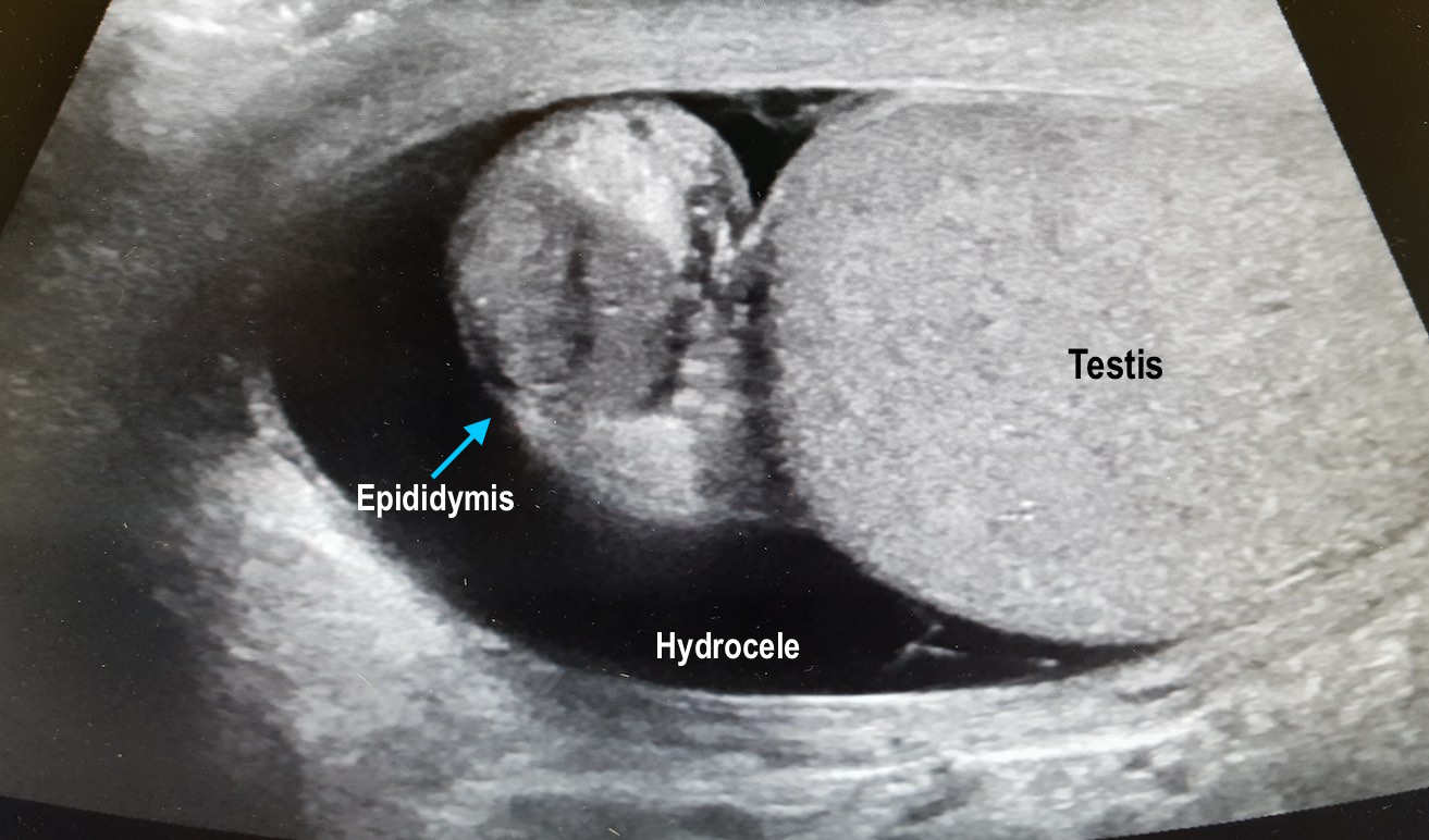
56 year-old male with history of hypertension presents with complaints of right scrotal swelling and pain. Denies any urinary symptoms, abdominal pain, nausea/vomiting or change in bowel habits or prior episodes. Temp was 99.0.
A scrotal ultrasound was done and an image of the right testis was seen (below). What's the diagnosis?
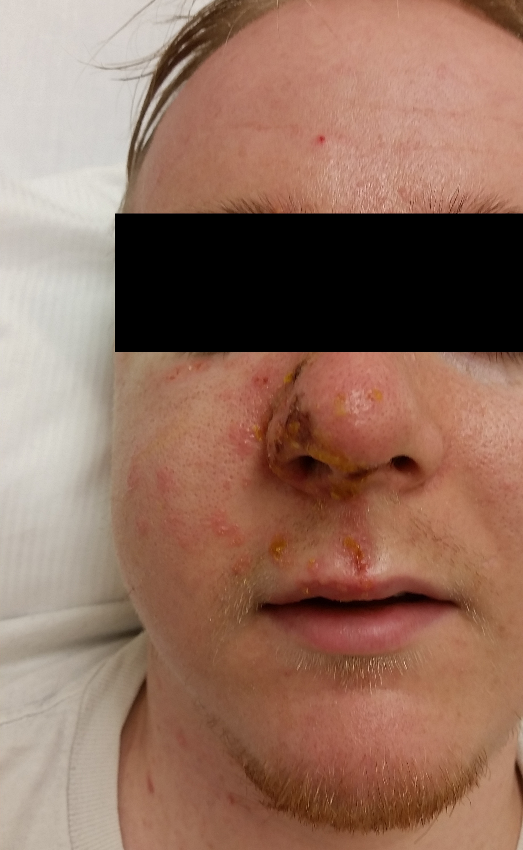
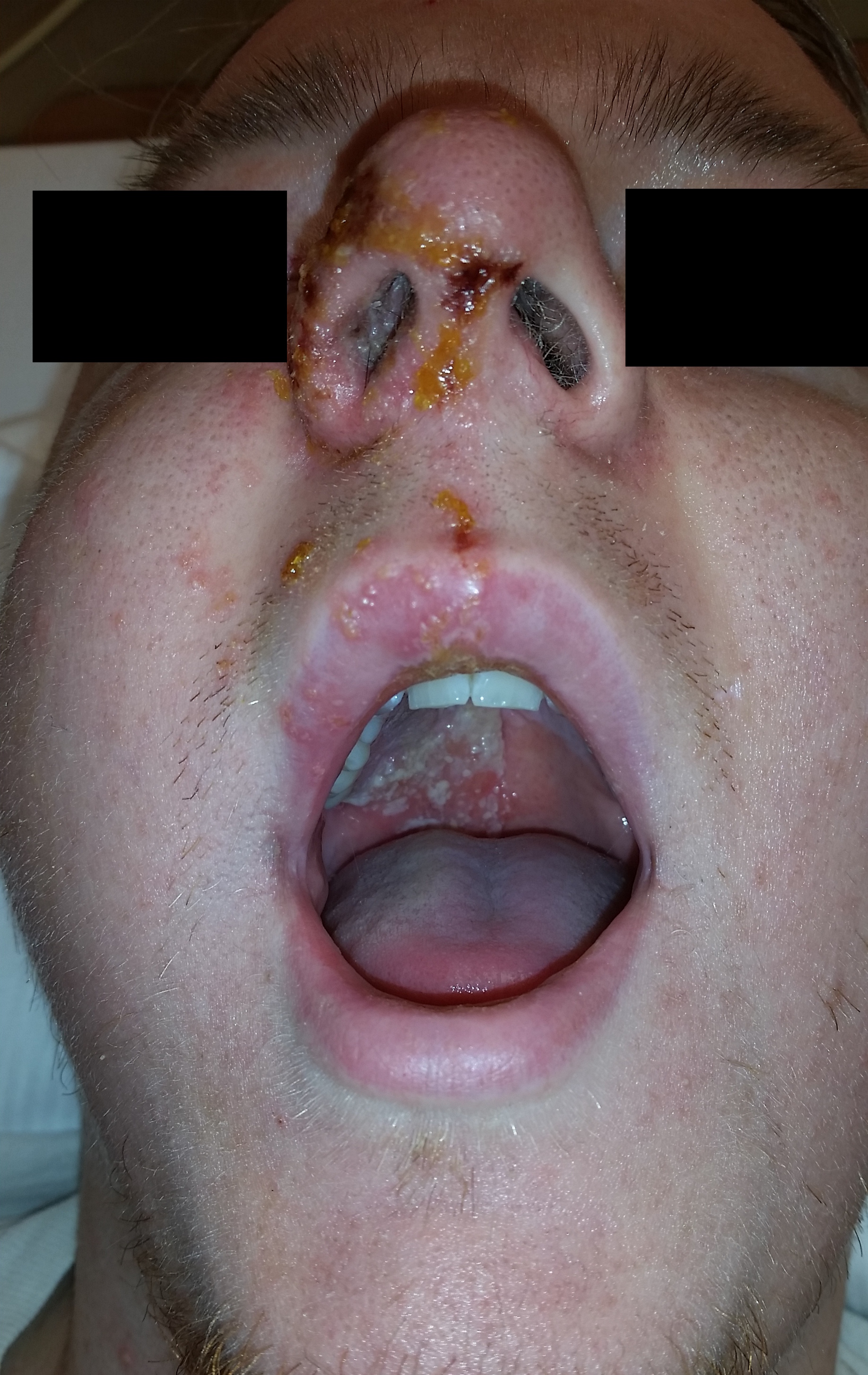
24-year-old male with a history of Wagner's Granulomatosis, currently on Cellcept (Mycophenolate Mofetil) and high dose prednisolone, presented with two days of sore throat, malaise and the lesions shown in the picture. What is the diagnosis?
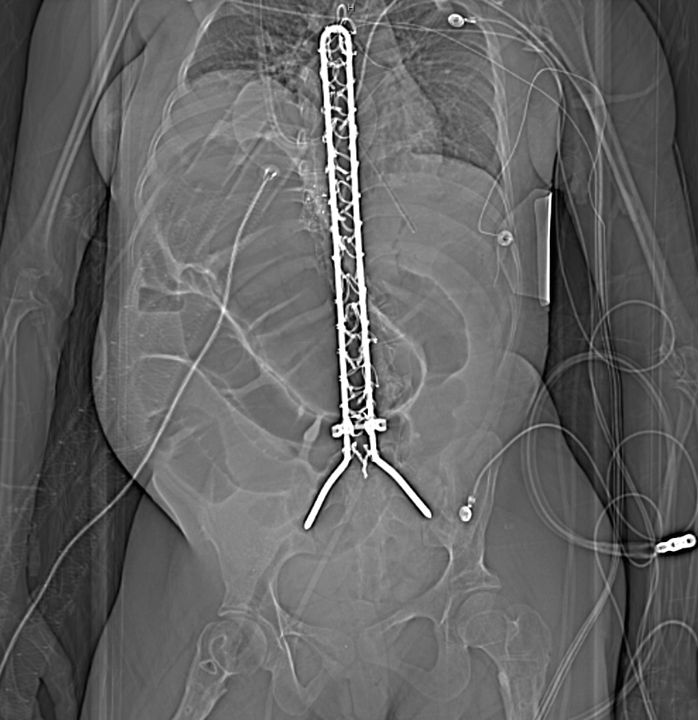
25 year-old female with hx of cerebral palsy with significant developmental delay, s/p G-tube who presented with acute hypoxic respiratory failure, hypotension and a distended, tense abdomen. A CT was done with the scout film below. What's the diagnosis?
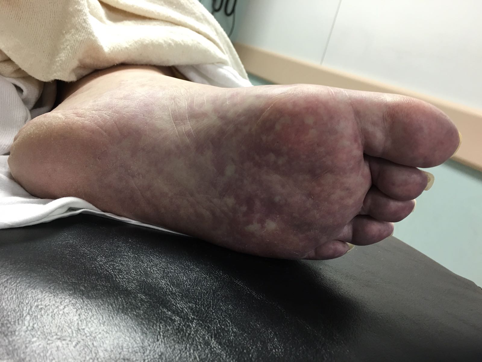
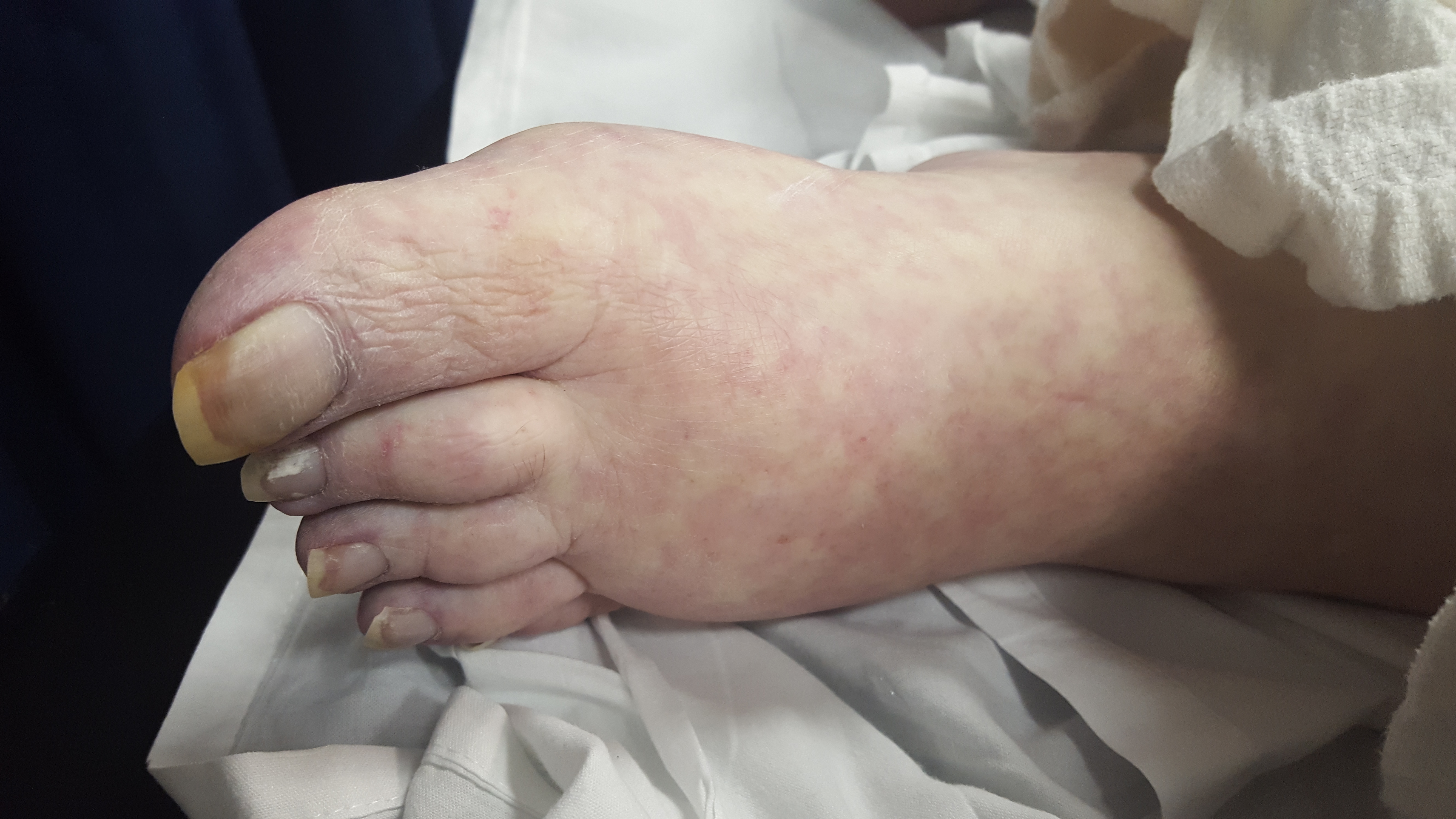
A 60 year-old man with history of atrial fibrillation, CAD presents with left lower leg/foot pain for a few days. His foot is seen below. What's the diagnosis?
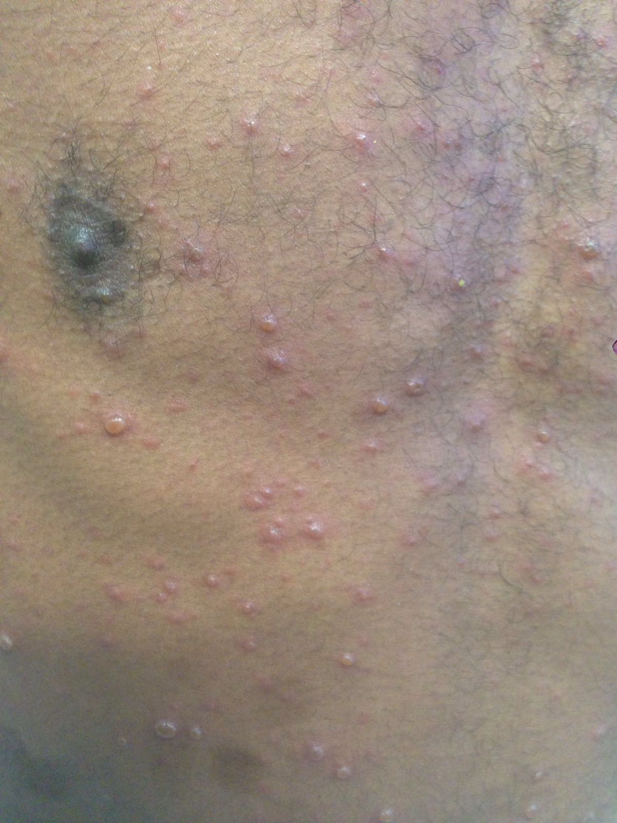
A 36-year-old male, who recently immigrated from Africa, presented to the ED with fever, rash, cough and shortness of breath. He was noted to be febrile to 39.0 C. The rash is disseminated but present mainly in his trunk as shown in the picture.
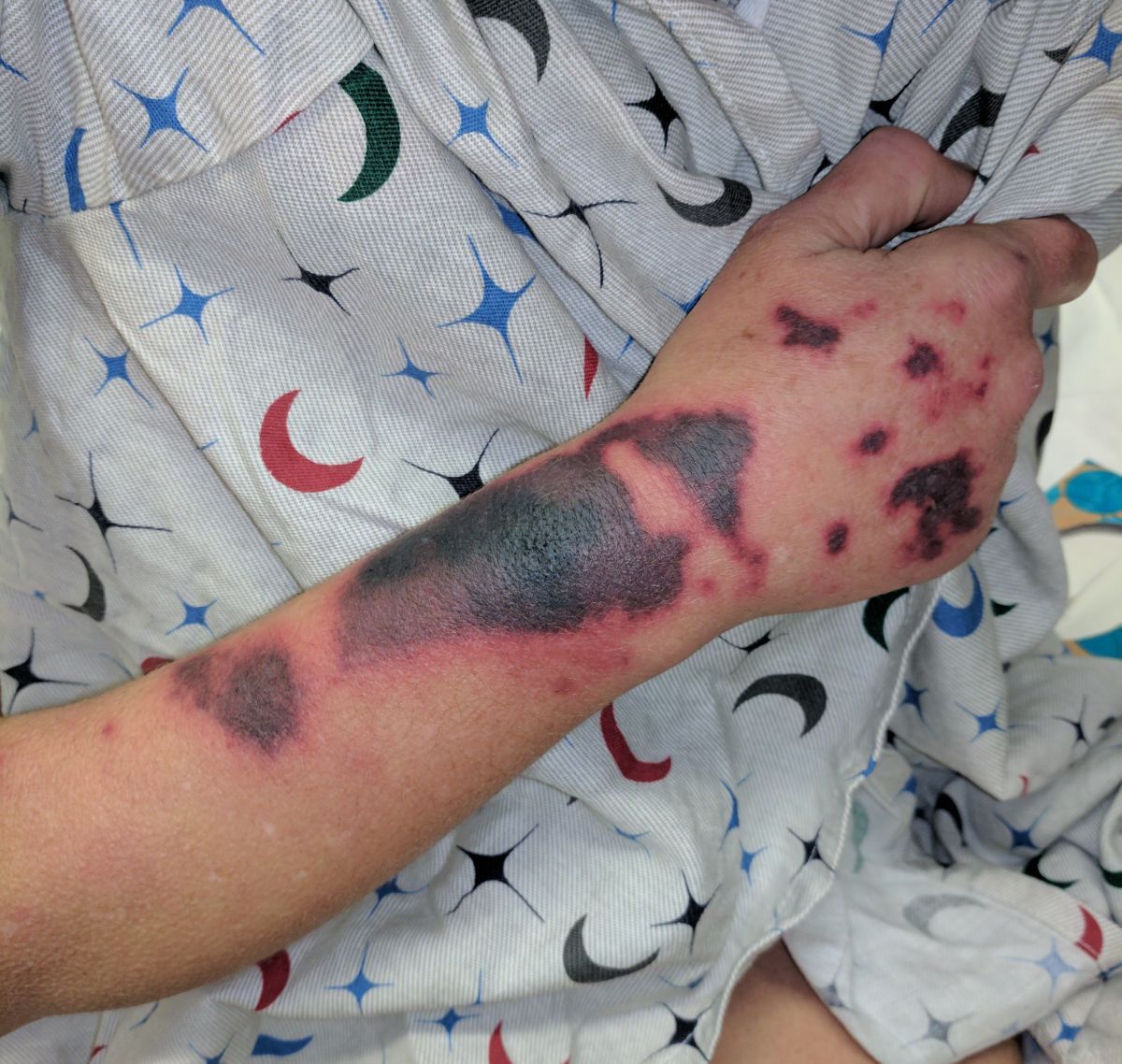
30 Year-old female presents to the ED for a rash. The rash started suddenly, mainly in her extremities and it is painful. The patient denied having fever or chills. Her past medical history is unremarkable. She admits to using cocaine frequently. The rash is shown in the picture.
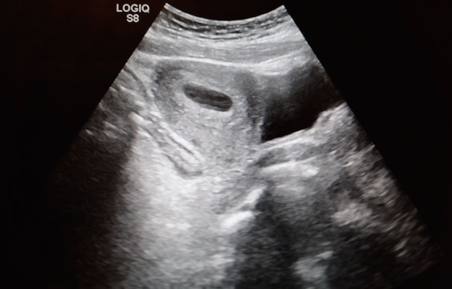
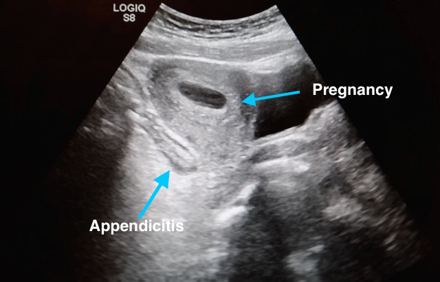
27 year-old G2P1 presents with 3 days of abdominal pain that is mostly suprapubic. Denies any urinary symptoms and vaginal bleeding. Physical examination reveals slight rebound in the right lower quadrant.
An ultrasound revealed the following. What's the diagnosis?
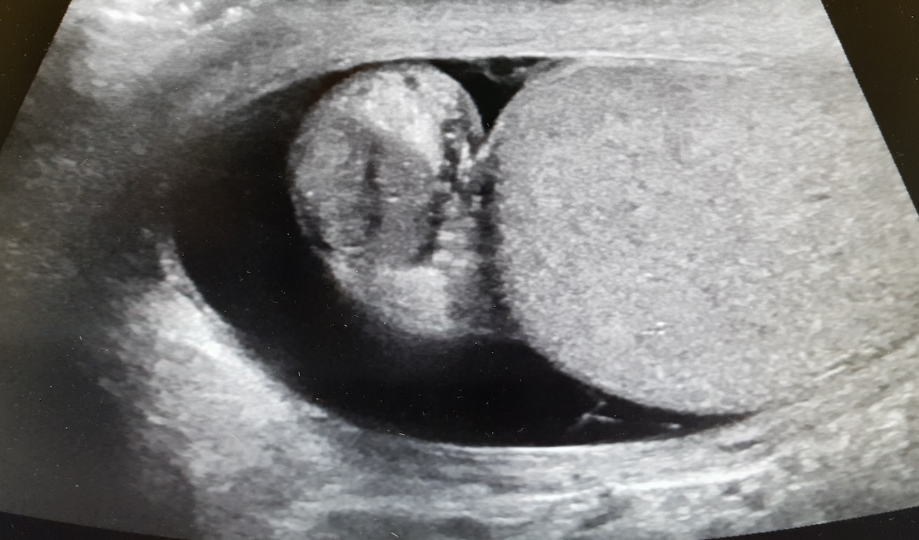
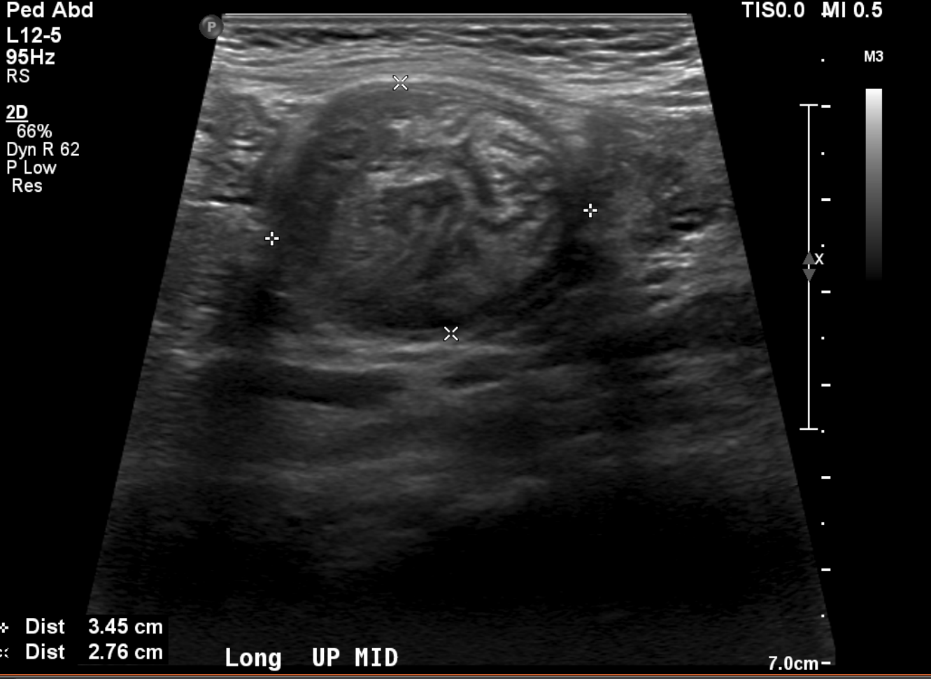
A 15 months old male with no past medical history, presenting with two days of decreased oral intake and decreased urine output. The exam was notable for minimal tenderness of abdomen. During an oral fluid challenge in the ED, the patient had a single episode of bilious vomiting. The ED physician ordered an ultrasound study and the results are shown below. What is the diagnosis?
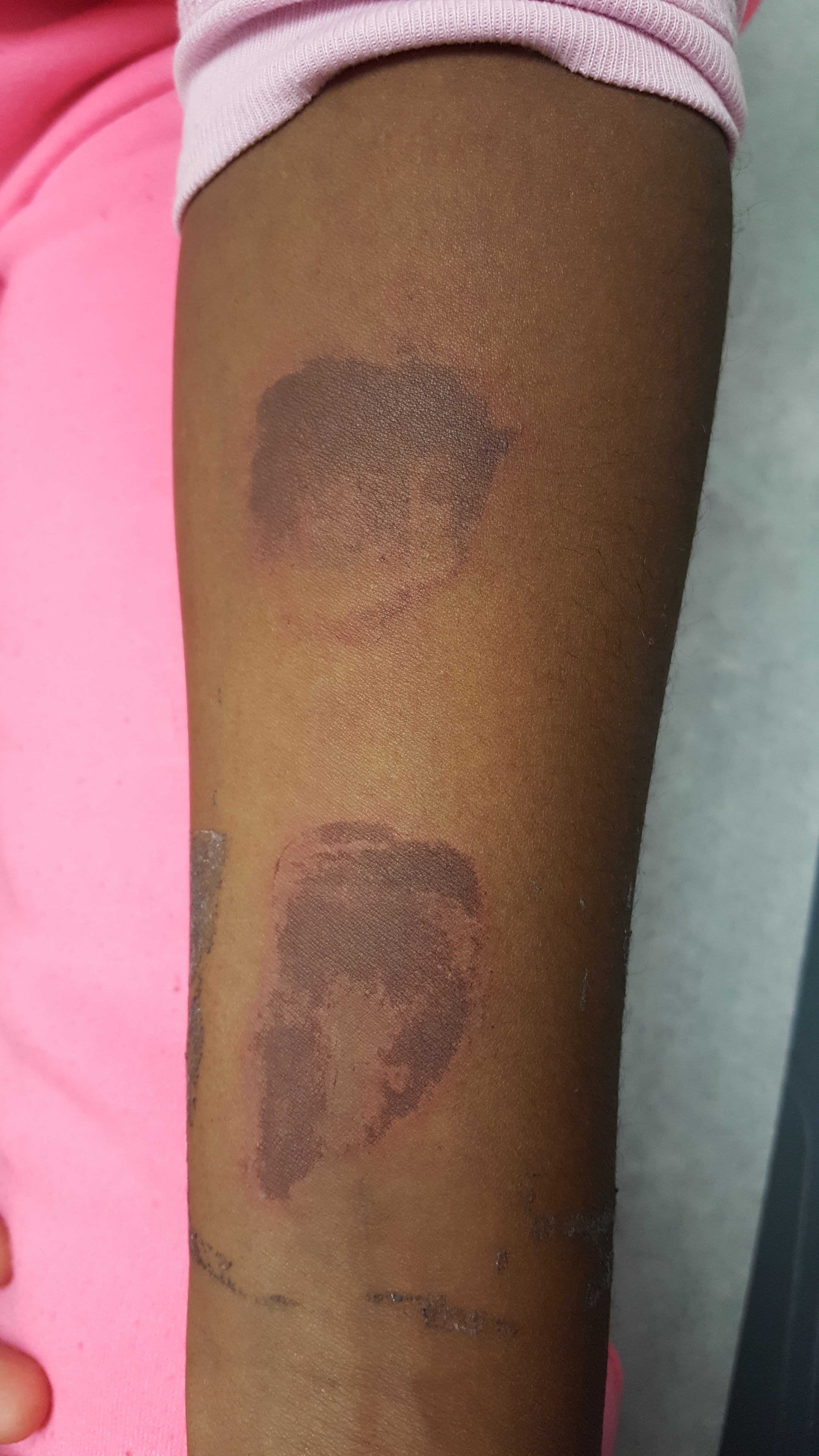
8 year-old female with no PMH who presents with concerns for "purple patches" popping up on her arm for 2-3 days. Stated that one appeared and then, the other one appeared 12 hours later. She denied any trauma whatsoever, history of easy bleeding/bruising and did feel safe at home. The rest of the review of systems was negative.
Patient said there was mild pain when the area was touched. The rest of the physical examination was normal.
What's the diagnosis? (Image below)
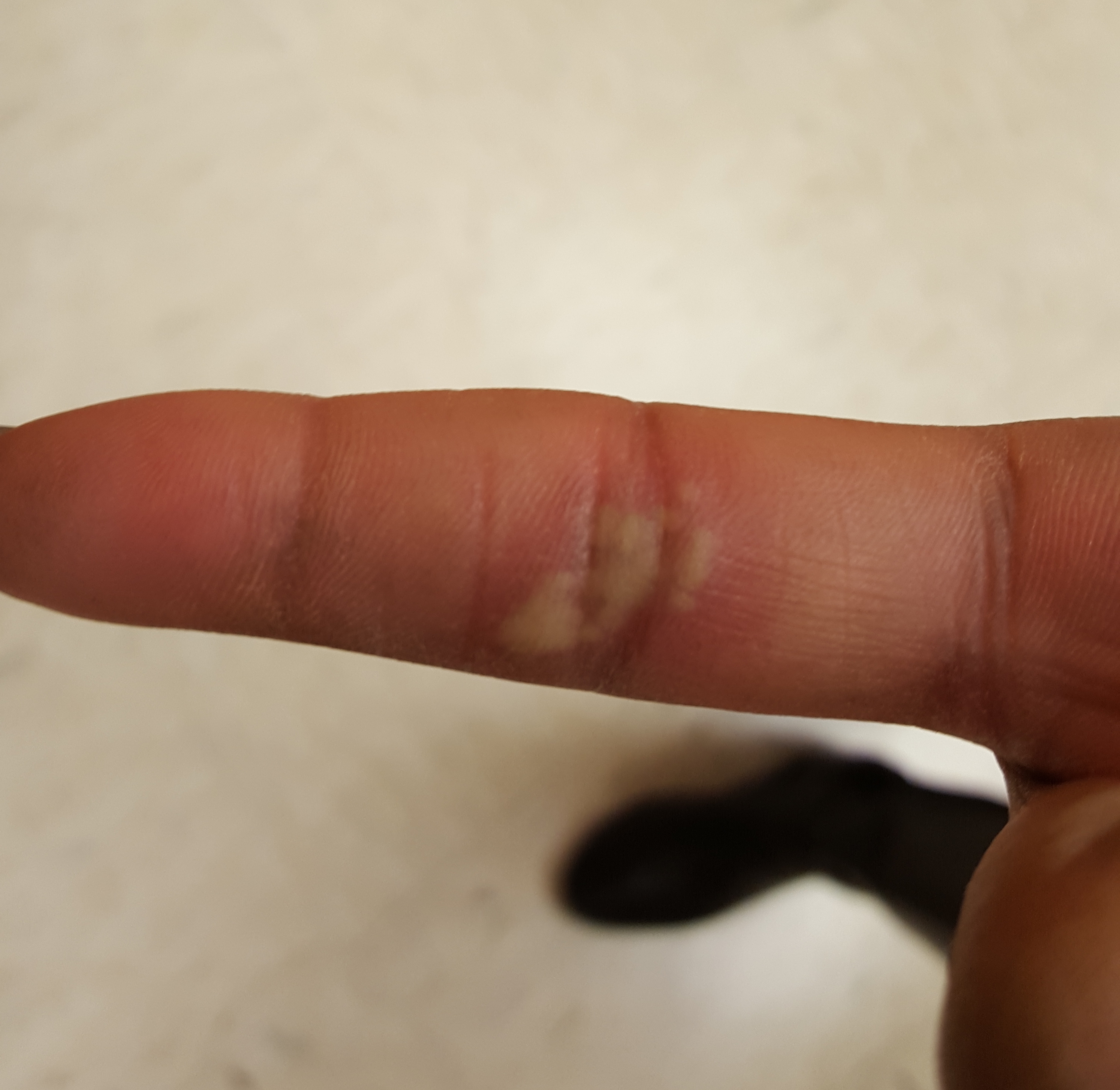
30 year old female presents with a painful finger for 1 week. Finger exam showed the following. What is the diagnosis ?


