Submitted by Dr. Lauren Rice
The summertime can be full of lots of fun activities (beach, fireworks, cookouts, and campfires) that can put children at risk of burns.
Burn depth classification:
1. Superficial (first-degree): red and blanching with minor pain, resolves in 5-7 days
2. Partial thickness (second-degree): red and wet with blisters, very painful, resolves in 2-5 weeks
Treatment: clean with soap and water twice daily, and apply silvadene wrap with gauze, kerlex
3. Full thickness (third-degree): dry and leathery without pain, no resolution after 5-6 weeks, may require graft
Treatment: wound debridement and dressings as above
Parkland formula: 4ml/kg/%TBSA in 1st 24 hours with 50% of total volume in 1st 8 hours
Calculate burn surface area:
-SAGE: free computerized burn diagram available at www.sagediagram.com
-Rule of Nines > 14 years old
-Rule of Palm <10 years old
Burn Center Referral
-Extent: partial thickness of >30% TBSA or full thickness of >10-20%
-Site: hands, feet, face, perineum, major joints
-Type: electrical, chemical, inhalation
Pathology at the umbilicus can manifest as inflammation, drainage, a palpable mass, or herniation.
Omphalitis - A cellulitis of the umbilicus. Mild cases often respond to local application of alcohol to clean the area, but due to the possibility of rapid progression and abdominal wall necrotizing fasciitis, admission for observation and IV antibiotics is usually warranted. Cover staph, strep, and GNRs.
Umbilical granuloma - As the umbilical ring closes and the cord sloughs off, granulation tissue formation is a normal part of umbilical epithelialization. There is sometimes an overgrowth of granulation tissue which can be treated once or twice with silver nitrate. Should the tissue not regress after a 1-2 treatments, the patient should be referred to pediatric surgery for excision and evaluation of other pathology (urachal or vitelline remnants).
Umbilical fistula - This is a patent vitelline duct and is characterized by persistent drainage that is bilious or purulent. A fistulogram using a small catheter and radio opaque dye can sometimes be helpful in determining the source of drainage (dye should be seen in the small bowel).
Umbilical polyp - Often confused with an umbilical granuloma with its glistening cherry red appearance, this is actually a vitelline duct remnant and contains small bowel mucosa. It does not regress with silver nitrate.
Vesicoumbilical fistula/sinus - The urachal versions of the umbilical fistula. This are a failure of complete closure of the urachus, resulting in persistent drainage of urine from the umbilicus, and infection (including recurrent UTIs). A fistulogram can be helpful for diagnosis.
Though an uncommon event, Drug-Induced Autoimmune thrombocytopenia occurs in a variety of drugs. Having recently diagnosed a patient that was receiving the "double-dose" bactrim for an MRSA abscess, it is worth mentioning the other drugs that have been reported to do it. Platelet count can go down to lethal levels and result in death due to the coagulopathy. Treatment is effective with platelets and no contraindication like in TTP.
Drugs that have been reported to do it:
abciximab, acetaminophen, amiodarone, amphotericin B
Carbamazepine, danazol, diclofenac, digoxin
Methyldopa, procainamide
Rifampin, trimethoprim-sulfamethoxazole, vancomycin
Acute Kidney Injury and Tumor Lysis Syndrome
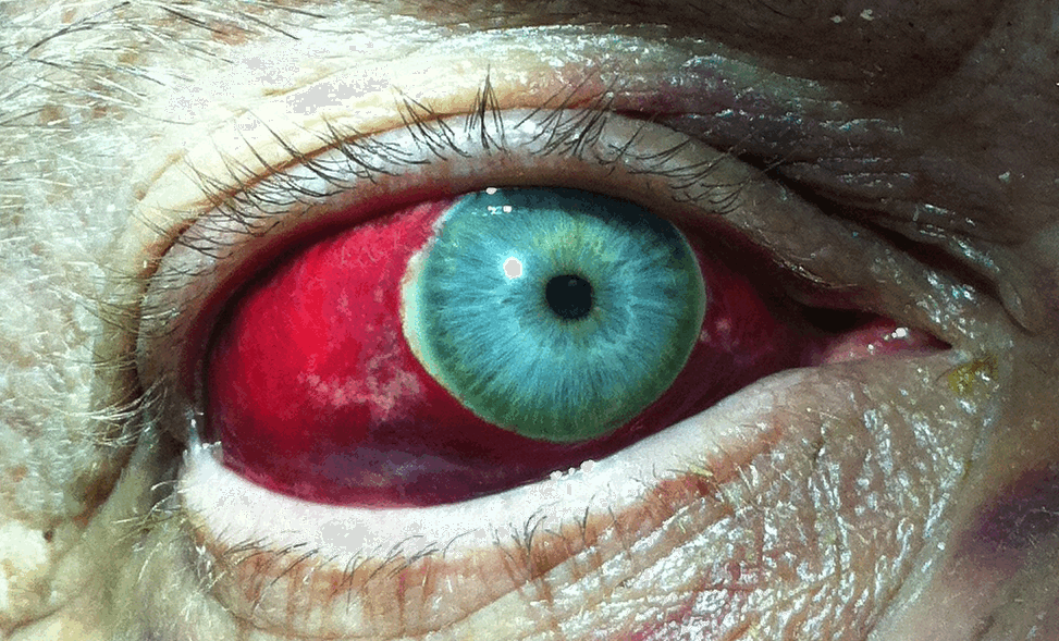
77 year old male presents to the Emergency Department one week after a motor vehicle crash in which he suffered minor facial injuries. He is now concerned because his eye looks like this. Diagnosis?
LBBB is defined by 3 criteria QRS >125msec, V1- QS or rS, and R wave peak time 60ms with no q wave in leads I, V5, V6
I am often asked whether physical activity has a positive or negative effect on the overall health of knee cartilage. The answer is unclear. Published data are conflicting.
What is known and generally agreed on:
1) Physical activity has been shown to facilitate cartilage development in children
2) Forced immobility (spinal cord injury) results in rapid cartilage loss
3) The medial knee compartment experiences significant mechanical loads during weight-bearing activity and is often the primary site of knee OA
A recent study attempted to answer whether 1) long-term (10yrs) participation in vigorous physical activity would benefit knee cartilage in healthy adults and 2) whether there were certain subgroups with asymptomatic preexisting structural knee changes which predict a harmful cartilage response to long-term physical activity.
Vigorous = activity generating sweating or SOB at least 20min 1/wk
Healthy older adults (mean age 57.8 yr) performing persistent vigorous physical activity had an increased risk (odds ratio 1.5) of worsening medial knee cartilage defects but not of a change in cartilage volume
In those w/ asymptomatic preexisting structural knee changes, there was worsening of cartilage defects (odds ratio 3.4) and a trend toward increased rate of loss of cartilage volume (again in the medial knee compartment)
Long-term effects of vigorous physical activity may depend on the preexisting health of the joint
Intussusception is the telescoping or prolapse of one portion of the bowel into an immediately adjacent segment.
Transplant patients are the norm now in the ED. Their drug lists are immense and are usually on some form of immunosuppression to prevent rejection of the transplanted organ. Two common medications are cyclosporine and tacrolimus. They share many adverse effects like hepatotoxicity, nephrotoxicity and hypertension. Here is the mechanism of action and some unique adverse effects to these powerful immunosuppressants (there are many more so be wary):
1) Cyclosporine - suppresses T-cell activation and growth. Unique toxicity - painful neuropathy of the fingertips and toes, cortical blindness
2) Tacrolimus - simiar to cyclosporine but actually hampers T-cell communication/signal transduction. Unique toxicity - can also cause cortical blindness but is also known to cause diabetes/hyperglycemiad
Two recently presented abstracts at the 2012 Society of Critical Care Medicine conference suggest that the combination of vancomycin and piperacillin-tazobactam may lead to acute kidney injury (AKI) in the critically ill. There may also be evidence to suggest that piperacillin-tazobactam alone increases the risk of AKI.
Both abstracts retrospectively compared patients who received either vancomycin alone or the combination of vancomycin and piperacillin-tazobactam. In both studies, the rates of AKI were significantly lower in patients treated with vancomycin alone as compared to patients receiving both vancomycin and piperacillin-tazobactam.
Bottom line: Although the current evidence does not support a change in our clinical practice, more prospective studies exploring this topic are necessary.
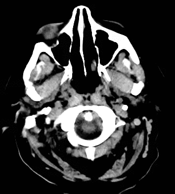
79 year old male with headaches, ataxia, falls, and difficulty urinating. What's the diagnosis?
For patients presenting to the ED with chest pain, we've been taught that “classic” or “typical” presentations for ACS (chest pressure with radiation to the left neck/jaw/shoulder/arm, dyspnea, diaphoresis, nausea, vomiting, lightheadedness) are most worrisome. Yet, many of the patients that present with typical symptoms end up having negative workups for ACS. What are the symptoms that truly predict ACS? Three major studies have demonstrated that the best predictors of ACS in patients presenting to the ED with chest pain are (not necessarily ranked in order):
1. chest pain that radiates to the arms, especially if the pain radiates bilaterally or to the right arm
2. chest pain associated with diaphoresis
3. chest pain associated with vomiting
4. chest pain associated with exertion
The description of the chest pain (e.g. "pressure" or "squeezing," etc.), the dyspnea, nausea, lightheadedness, and pain at rest were, surprisingly, not helpful at predicting ACS.
The simple takehome point is the following: always ask your patient with chest pain if the pain radiates, if there was associated diaphoresis, if there was associated vomiting, and if the pain is associated with exertion. If the answers to any of these 4 questions is "yes," think twice before labeling the patient with a non-ACS diagnosis.
Contrast Allergy:
Many patients will report that they have a allergy to iodinated contrast by saying that they are allergic to iodine
Iodine, itself, is not an allergen and is a required element for thyroid homrone production. Plus could you imagine the hordes of people that would be having allergic reactions everyday when they add salt to their french fries. Our EDs would be completely swamped.
A recent meta-analysis by Drs. Schabelman and Witting also showed the following:
As we enter Crab eating season in Maryland, lets stop giving shellfish a bad name. A patent with any allergy is at increased risk, but shellfish is no higher a risk than those allergic to Strawberries.
Definition: Fracture of the humerus just proximal to the epicondyles.
Use the Measured Sodium Concentration!
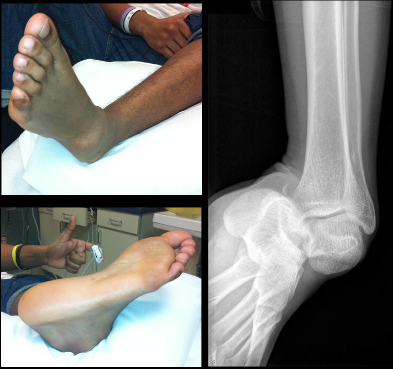
19 year-old male presents with L ankle pain and obvious deformity after jumping out of a window and landing on his inverted foot. What's the diagnosis?
New studies are utilizing mild therapeutic hypothermia as a treatment option in cardiogenic shock. These studies have reported improved circulatory support, an increase in systemic vascular resistance, and reduction in vasopressor use which ultimately may result in lower cardiac oxygen consumption. The preliminary results suggest that mild therapeutic hypothermia could be a therapeutic option in hemodynamically unstable patients independent of current recommendations which support its use in cardiac arrest survivors.
• Wedge compression fractures
http://jbjs.org/data/Journals/JBJS/855/JBJA0851224560G02.jpeg
It may not be necessary to give oral vitamin K to patients that are not bleeding that have INRs between 4.5 and 10.
Patients who were supratherapeutic on warfarin were randomized to vitamin K 1.25 mg (n=355) versus placebo (n=369).
In the 90 days after enrollment, 15.8% of patients allocated to vitamin K and 16.3% allocated to placebo had a bleeding event. Major bleeding events occurred in 9 patients in the vitamin K group and 4 in the placebo.
Thromboembolic events occurred in 1.1% of patients in the vitamin K group, compared to 0.8% of patients in the placebo group. An equal number of patients died in each group (n=7).