Category: Orthopedics
Keywords: overuse injury, wrist (PubMed Search)
Posted: 5/25/2023 by Brian Corwell, MD
Click here to contact Brian Corwell, MD
Intersection syndrome
Intersection syndrome is an overuse injury of the forearm.
Pain is located approximately 2 finger breaths (4cm) proximal to the wrist joint.
https://www.sportsmedreview.com/wp-content/uploads/2020/11/intersectionsyndrome.png
Mechanism: friction is caused by repetitive wrist extension activities
Commonly: Rowing, skiing, tennis, canoeing and weightlifting
Friction may cause crepitus with finger/wrist extension.
Tenderness, mild swelling may be present
Category: Visual Diagnosis
Keywords: C Spine, osteomyelitis, (PubMed Search)
Posted: 5/25/2023 by Robert Flint, MD
(Updated: 2/2/2026)
Click here to contact Robert Flint, MD
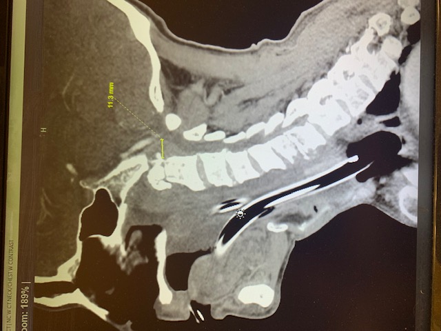
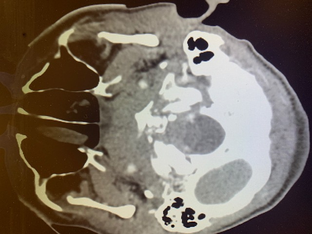
Neck pain and trouble swalowing. No trauma.
Osteomyelitis of the cervical spine is the least common location to find spinal infection. “Osteomyelitis of the cervical spine is a rare disease, representing only 3% – 6% of all cases of vertebral osteomyelitis. In contrast with other locations of spinal infections, osteomyelitis of the cervical spine can be a much more dramatic and rapidly deteriorating process, leading to early neurologic deficit”.
Note the bony destruction as well as the abscess labeled on the lateral view.
Category: Administration
Keywords: patient experience, clinician wellbeing (PubMed Search)
Posted: 5/24/2023 by Mercedes Torres, MD
Click here to contact Mercedes Torres, MD
Clinician Well-Being and the Patient Experience
Did you know that most patient experience responses are overwhelmingly positive? Rather than focusing all our attention on the bad, let’s focus on the good to promote clinician well-being. See below for a few key points from a recent study on this:
Consider emphasizing positive patient experiences when providing feedback to emergency physicians. It will promote clinician well-being and help improve performance in your practice.
Dudley J and Lee TH. Patient Experience and Clinician Well-Being Aren’t Mutually Exclusive. Harvard Business Review. Published online at hbr.org, July 18, 2022.
Category: Critical Care
Keywords: Intubation, Trauma, Cervical Spine, Laryngoscopy (PubMed Search)
Posted: 5/23/2023 by Mark Sutherland, MD
Click here to contact Mark Sutherland, MD
Ability to move the head and neck freely can be clutch in endotracheal intubation, so in patients such as certain trauma patients who may have c-spine instability and need to be immobilized, it's all the more important to choose the optimal intubation approach to maximize success and minimize head movement.
Choi et al recently published a study in Anesthesia looking at:
-Video laryngoscopy with a standard geometry Mac blade
vs
-Fiberoptic intubation
as the initial method for intubating patients in c-collars about to undergo spinal surgery. This is an interesting contrast between two extremes, as standard geometry is the most "traditional" approach, whereas fiberoptic is kind of the opposite end of the spectrum, jumping to a more advanced method which might be more flexible (no pun intended) but also introduces new complexities.
All outcomes actually favored standard geometry VL over fiberoptic, including first pass success (98% vs 91%), time to intubation (50s vs 81s) and need for additional airway maneuvers (18% vs 56%). There was no difference in complication rates, although a bigger study might be needed to find rare complications (this study had 330 patients).
In my opinion, it's unfortunate they didn't include hyperangulated VL, as it would be interesting to see how this approach compares. Personally I think of hyperangulated VL in these patients as a nice blend of the two methods, bringing the familiarity and speed of typical VL intubation, but often requiring less neck movement like fiberoptic.
Bottom Line: This study does not support a fiberoptic first approach to intubating patients with cervical spine instability. In fact, it may cause harm.
Choi S, Yoo HK, Shin KW, Kim YJ, Yoon HK, Park HP, Oh H. Videolaryngoscopy vs. flexible fibrescopy for tracheal intubation in patients with cervical spine immobilisation: a randomised controlled trial. Anaesthesia. 2023 May 5. doi: 10.1111/anae.16035. Epub ahead of print. PMID: 37145935.
Category: Trauma
Keywords: circulation, trauma, hemorrhage, atls (PubMed Search)
Posted: 5/20/2023 by Robert Flint, MD
(Updated: 2/2/2026)
Click here to contact Robert Flint, MD
It is time to abandon the ABC's that ATLS teaches and move to hemorhage control (circulation) as well as resucitation before we deal with airway in the majority of trauma patients. Tounriquets save lives. Pelvic binders save lives. Blood transfusion (whole blood) saves lives. Poisitive presssure ventilation, sedativies, and decreasing sympathetic drive in hypoternsive patients makes their hypotension worse.
Please consider changing to a CAB approach to the hyhpotensive trauma patient.
Ferrada P, Dissanaike S. Circulation First for the Rapidly Bleeding Trauma Patient—It Is Time to Reconsider the ABCs of Trauma Care. JAMA Surg. Published online May 17, 2023. doi:10.1001/jamasurg.2022.8436
Category: Quality Assurance/Quality Improvement
Keywords: Ct scan, abdominal pain, IV contrast, diagnosis (PubMed Search)
Posted: 5/20/2023 by Robert Flint, MD
(Updated: 2/2/2026)
Click here to contact Robert Flint, MD
Does IV contrast help to make the diagnosis in ED abdominal pain patients undergoing CT scan? The authors of this study tried to answer that question. This study was a retrospective diagnostic accuracy study looking at contrast enhanced vs. non-enhanced images in 201 consecutive ED patients. The study demographics were:
“There were 201 included patients (female, 108; male, 93) with a mean age of 50.1 (SD, 20.9) years and mean BMI of 25.5 (SD, 5.4).”
The study found: “Unenhanced CT was approximately 30% less accurate than contrast-enhanced CT for evaluating abdominal pain in the ED.”
This study is limited by the small size, the overwhelming female to male inclusion, the reliance on radiology reading as the gold standard of pathology, and the retrospective nature. It does, however, show that there is a need for further study and at this time giving IV contrast has limited down side and potentially improves diagnostic accuracy of abdominal CT scans.
Shaish H, Ream J, Huang C, et al. Diagnostic Accuracy of Unenhanced Computed Tomography for Evaluation of Acute Abdominal Pain in the Emergency Department. JAMA Surg. Published online May 03, 2023. doi:10.1001/jamasurg.2023.1112
Category: Pediatrics
Keywords: IV, EMS, transfer, pediatrics (PubMed Search)
Posted: 5/19/2023 by Jenny Guyther, MD
(Updated: 2/2/2026)
Click here to contact Jenny Guyther, MD
Mangus CW, Canares T, Klein BL, et al. Interhospital Transport of Children With Peripheral Venous Catheters by Private Vehicle: A Mixed Methods Assessment. Pediatr Emerg Care. 2022;38(1):e105-e110. doi:10.1097/PEC.
Category: Misc
Keywords: EMS, Alternate destinations, pediatric, EMS, reduce transport times (PubMed Search)
Posted: 5/17/2023 by Jenny Guyther, MD
(Updated: 2/2/2026)
Click here to contact Jenny Guyther, MD
Ward CE, Singletary J, Campanella V, Page C, Simpson JN. Caregiver Perspectives on Including Children in Alternative Emergency Medical Services Disposition Programs: A Qualitative Study [published online ahead of print, 2023 May 5]. Prehosp Emerg Care. 2023;1-9. doi:10.1080/10903127.2023.
Category: Critical Care
Posted: 5/16/2023 by Mike Winters, MBA, MD
(Updated: 2/2/2026)
Click here to contact Mike Winters, MBA, MD
Bicarbonate Use for Lactic Acidosis?
Wardi G, et al. A review of bicarbonate use in common clinical scenarios. J Emerg Med. 2023; online ahead of print.
Category: Administration
Keywords: POCUS, Cardiac Arrest, Arterial Doppler (PubMed Search)
Posted: 5/15/2023 by Alexis Salerno Rubeling, MD
(Updated: 2/2/2026)
Click here to contact Alexis Salerno Rubeling, MD
Did you know that you can use the linear probe with pulse wave (PW) doppler over the femoral artery to look for a pulse during CPR pauses?
Well, the researchers of this article took this skill one step further to evaluate if they could use the femoral artery PW doppler while CPR was in progress to look for signs of a pulse.
The authors found that:
- pulsations due to compressions were organized with uniform pulsations.
- when there was also native cardiac activity, the pulsations were nonuniform and may have an irregular cadence.
Although there were several limitations, Arterial doppler was 100% specific and 50% sensitive in detecting organized cardiac activity during active CPR.
Take Home Point: Take a look at your arterial doppler for signs of organized cardiac activity during a resuscitation.
Reference: Gaspari RJ, Lindsay R, Dowd A, Gleeson T. Femoral Arterial Doppler Use During Active Cardiopulmonary Resuscitation. Ann Emerg Med. 2023 May;81(5):523-531. doi: 10.1016/j.annemergmed.2022.12.002. Epub 2023 Feb 7. PMID: 36754697.
Category: Trauma
Keywords: hypoxia, delayed sequence, RSI, Ketamine, succinylcholine (PubMed Search)
Posted: 5/7/2023 by Robert Flint, MD
(Updated: 2/2/2026)
Click here to contact Robert Flint, MD
Delayed sequence intubation can be valuable in the agitated, combative trauma patients who will not tolerate pre-intubation pre-oxygenation. We know peri-intubation hypoxia leads to significant morbidity and mortality. DSI offers us an option to avoid peri-inubation hypoxia.
This study randomized 200 trauma patients into a rapid induction group (Ketamine followed immediately by succinylcholine with immediate intubation) vs. delayed induction group (Ketamine followed by a 3-minute oxygenation period followed by succinylcholine, followed by intubation). The authors found: “Peri-intubation hypoxia was significantly lower in group DSI (8 [8%]) compared to group RSI (35 [35%]; P = .001). First-attempt success rate was higher in group DSI (83% vs 69%; P = .02).”
Anjishnujit Bandyopadhyay 1, Pankaj Kumar, Anudeep Jafra, Haneesh Thakur, Laxmi Narayana Yaddanapudi, Kajal Jain Peri-Intubation Hypoxia After Delayed Versus Rapid Sequence Intubation in Critically Injured Patients on Arrival to Trauma Triage: A Randomized Controlled Trial. Anesth Analg. 2023 May 1;136(5):913-919. doi:10.1213/ANE.0000000000006171. Epub 2023 Apr 14.
Category: Orthopedics
Keywords: Baker's cyst, knee, effusion (PubMed Search)
Posted: 5/13/2023 by Brian Corwell, MD
Click here to contact Brian Corwell, MD
A Baker's cyst is a common incidental finding on ultrasound reports and bedside physical exam.
Clinically, these cysts are commonly found in association with intra-articular knee disorders. Most commonly: osteoarthritis, RA and tears of the meniscus.
Sometimes Baker's cysts are a source of posterior knee pain.
In an orthopedic clinic setting, Baker’s cysts are frequently discovered on routine MRI in patients with symptomatic knee pain. They tend to occur in adults from ages 35 to 70.
Over 90% of Baker’s cysts are associated with an intraarticular knee disorder. While most frequently associated with OA and meniscal tears, other knee pathologies that have been associated include inflammatory arthritis and tears of the anterior cruciate ligament.
DDX: DVT, cystic masses (synovial cyst), solid masses (sarcoma) and popliteal artery aneurysms.
Based on cadaveric studies, a valvular opening of the posterior capsule, proximal/medial and deep to the medial head of the gastrocnemius is present in approximately 50% of healthy adult knees.
Fluid flows in one way from knee joint to cyst and not in reverse. This valve allows flow only during knee flexion as it is compressed shut during extension due to muscle tension.
Most common patient complaint is that of the primary pathology, meniscal pain for example. At times, symptoms related to the cyst are likely due to increasing size as they may report fullness, achiness, stiffness.
In one small study, the most common symptoms were 1) popliteal swelling and 2) posterior aching. Patients may complain of loss of knee flexion from an enlarged cyst that can mechanically block full flexion.
If the Baker cyst is large enough the clinician will feel posterior medial fullness and mild tenderness to palpation. The cyst will be firm and full knee extension and softer during the flexion (Foucher’s sign).
This may help with differentiation from other popliteal masses (hematoma, soft tissue tumor, popliteal artery aneurysm).
With cyst rupture, severe pain can simulate thrombosis or calf muscle rupture, (warmth, tenderness, and erythema). A ruptured cyst can also produce bruising, which may involve the posterior calf starting from the popliteal fossa and extending distally towards the ankle.
Treatment: initial treatment for symptomatic Baker cysts is nonoperative unless vascular or neural compression is present (very unlikely)
Treatment involves physical therapy to maintain knee flexibility. A sports medicine physician may perform an intraarticular knee corticosteroid injection as this has been found to decrease size and symptoms of cysts in two-thirds of patients.
For patients that fail above, refer for surgical evaluation. Inform patients that they are not undergoing ED drainage of this symptomatic cyst due to the extremely high rate of recurrence which, as a result of the ongoing presence of the untreated intraarticular pathology, results in the recurrent effusion.
Category: Pharmacology & Therapeutics
Keywords: contraception (PubMed Search)
Posted: 5/11/2023 by Ashley Martinelli
(Updated: 2/2/2026)
Click here to contact Ashley Martinelli
Emergency contraception is highly effective at preventing unwanted pregnancies and has been on the market for 20+ years.
Levonogestrel (LNG) 1.5 mg PO x 1 dose (OTC Available)
Ulipristal acetate (UPA) 30 mg PO x 1 dose (Requires RX)
Original studies with LNG was estimated to prevent up to 80% of expected pregnancies. In the subsequent trials that brought UPA to the market and compared the two medications, LNG prevented 69% (95% CI, 46-82%) and 52.2% (95% CI, 25.1-69.5%).
While pregnancy rates are low with both options there is concern with patients of higher weight/BMI that the effectiveness of levonorgestrel decreases as weight rises. One large study of over 1700 patients specifically noted that a weight > 75 kg was associated with up to 6.5% pregnancy rate (95% CI 3.1-11.5) compared to 1.4% (95% CI 0.5-3.0) in patients weighing 65-75 kg. Patients weighing > 85 kg had similarly high rates at 5.7% (95% CI 2.9-10.0).
The cost difference is minimal between products, especially when considering costs associated with treatment failures and subsequent need for care- the largest difference is with respect to access as LNG is OTC and UPA requires an RX. Either can be administered in an ED setting as long as they are on formulary.
ACOG also recommends that ulipristal be utilized for it higher overall efficacy compared to levonorgestrel.
Consider:
For patients above 75 kg, ulipristal can be used as first line emergency contraception for up to 5 days following unprotected intercourse.
Patients < 75 kg and < 72 hours following unprotected intercourse can use levonorgestrel or ulipristal as an appropriate emergency contraception method.
Patients < 75 kg and 72-120 hours following unprotected intercourse should use ulipristal due to its efficacy beyond 72 hours.
Kapp N, Abitbol JL, Mathe H, et al. Effect of body weight and BMI on the efficacy of levonogestrel emergency contraception. Contraception. 2015;91(2):97-104.
Emergency contraception. Practice Bulletin No. 152. American College of Obstetricians and Gynecologists. Obstet Gynecol 2015;126:e1–11
Category: Gastrointestional
Keywords: cannabis hyperemesis syndrome (PubMed Search)
Posted: 5/10/2023 by Neeraja Murali, DO, MPH
Click here to contact Neeraja Murali, DO, MPH
Bottom Line: With the increasing acceptance and legalization of marijuana and its derivatives, emergency departments have seen an increase in patients with cannabis hyperemesis syndrome (CHS). In this patient population, when other pathologies have been excluded, consider droperidol (0.625 mg – 2.5 mg) or haloperidol (0.05 mg/kg or 0.1 mg/kg) for management of symptoms.
Two separate articles were reviewed for this pearl. One is a systematic review of existing literature, and the other is a randomized controlled trial.
The systematic review examined 17 existing studies, including case reports, RCTs, retrospective studies, and other systematic reviews. This included adults aged 18-85 who were using recreational or medicinal cannabinoids. There was a consensus that cessation of cannabinoid use is the best way to alleviate symptoms of CHS. Other options discussed include:
As mentioned above, the HaVOC study examined various doses of haloperidol versus odansetron. This randomized controlled trial was triple blinded and had three groups: haloperidol 0.05 mg/kg or 0.1 mg/kg or odansetron 8 mg IV. The outcome of interest was reduction in abdominal pain and nausea at two hours after treatment. Either dose of haloperidol was found to be superior to odansetron, with improvements in pain and nausea (54% versus 29%; 95% CI -16% to 59%), and less use of rescue antiemetics (31% versus 59%, with 95% CI -61% to 13%). Haloperidol also resulted in shorter ED length of stay (3.1 h vs 5.6 h, 95% CI 0.1-5.0 h, p=0.03). However, 2 patients in the high dose haloperidol group had dystonic reactions precipitating return visits. The study does not specifically discuss differences in outcomes between the high and lower dose haloperidol groups.
Neither paper discussed the best alternatives when QTc prolongation is of concern. Clinicians should use their best judgment and the available information when deciding on a treatment option.
Senderovich H, Patel P, Jimenez Lopez B, Waicus S. A Systematic Review on Cannabis Hyperemesis Syndrome and Its Management Options. Med Princ Pract. 2022;31(1):29-38. doi:10.1159/000520417
Ruberto AJ, Sivilotti MLA, Forrester S, Hall AK, Crawford FM, Day AG. Intravenous Haloperidol Versus Ondansetron for Cannabis Hyperemesis Syndrome (HaVOC): A Randomized, Controlled Trial. Ann Emerg Med. 2021;77(6):613-619. doi:10.1016/j.annemergmed.2020.08.021
Category: Critical Care
Keywords: OHCA, Critical Care, Whole Body CT, Post Cardiac Arrest Care (PubMed Search)
Posted: 5/9/2023 by Lucas Sjeklocha, MD
Click here to contact Lucas Sjeklocha, MD
Just scan ‘em? Should everyone with unexplained out-of-hospital cardiac arrest get whole-body CT/CTA?
Background: Determination of the cause and subsequent management of out-of-hospital cardiac arrest is clinically challenging in those patients who survive to hospital admission without a clear diagnosis. CT imaging is often used to ascertain the cause of an arrest, find potentially intervenable etiologies, and assess for neurological injury but this practice and diagnostic yield are inconsistent and not well studied.
Study and Methods: The CT FIRST study is a single center cohort study using head-to-pelvis contrasted triple phase CT within 6 hours for cardiac arrest without obvious cause (sudden death CT or SDCT) studied in a before and after manner compared to usual care to determine the influence of early pan CT on diagnostic yield and outcomes. The primary outcome was diagnostic yield following SDCT and secondary outcomes include time to diagnosis of “time critical” findings and survival to discharge. 104 patients undergoing SDCT were compared to 143 historical controls after study implementation. Patients deemed to have a clear cause or are too unstable for CT were among exclusions.
Results: For the primary outcome of diagnostic yield: 92% of SDCT cohort received a separately adjudicated diagnosis for the arrest compared to 75% of the control cohort (p = 0.001). With time to such diagnosis of 3.1hrs in SDCT versus 14.1hrs of controls, with 39% versus 17% being made by CT. Time critical diagnoses including MI, PE, aortic dissection, pneumonia, embolic or hemorrhagic CVA and abdominal catastrophe were identified in 32% versus 24% (non significant) of the cohorts with delay greater than 6hrs to diagnosis reported in 12% in SDCT versus 62% in usual care (p=0.001).
There was no difference in survival to hospital discharge and no difference in safety measures and no evaluation reporting changes to and timing of patient managements.
The SDCT cohort had 100% scan rate compared to usual care where 81% received early head CT with chest CT and abdominal CT done in 36% and 18%, respectively. Notably there were no CT reported diagnoses that were later reversed on adjudication in either cohort. The planned economic and resource analysis was not reported in this study.
Discussion: There was a notable increase in diagnostic yield based on the study design with faster time to potentially time sensitive diagnoses. There were, however, no differences in mortality and it was not clear the degree to which these diagnoses influenced patient management given the limited numbers in this study and diverse set of diagnoses associated with cardiac arrest. Like previous studies of selective versus whole body CT in trauma populations, the increased diagnostic yield was not associated with reduced mortality or reported changes in management. The yield numbers suggest increased confidence by exclusion as much as positive findings of the cause. As always, the caveats of a relatively small single center before-and-after cohort study apply.
An interesting twist is that no CT diagnosis pointing to the cause of the arrest was reversed on subsequent review, this may speak to the accuracy of modern CT and radiology interpretation, but I sometimes worry that this can also be reflective of diagnostic fixation, especially with “objective” tests, as well as nihilism about the utility of clinical diagnosis.
That said, non-selective CT has many potential benefits for many critically ill and unexaminable populations with diagnostic uncertainty, as demonstrated here, which must be balanced against risks of intrahospital transport and of resource utilization as we do not yet have clear data that patients benefit from the practice despite increased diagnostic yield.
Kelley R.H. Branch, M.O. Gatewood, P.J. Kudenchuk et al., Diagnostic yield, safety, and outcomes of Head-to-pelvis sudden death CT imaging in post arrest care: The CT FIRST cohort study, Resuscitation, https://doi.org/10.1016/j. resuscitation.2023.109785
Category: Cardiology
Keywords: Posterior MI, ECG (PubMed Search)
Posted: 5/8/2023 by Leen Alblaihed, MBBS, MHA
Click here to contact Leen Alblaihed, MBBS, MHA
52 yo M with chest pain and shortness of breath, ECG as shown, do you activate cath lab?

The posterior descending artery (PDA) supplies the posterior third of the interventricular septum, including the posterior and inferior walls of the left ventricle. The vessel most commonly originates from either the right coronary artery (right dominant), left circumflex artery (left dominant), or both (codominant).
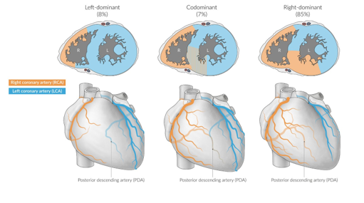
Posterior MI frequently occurs as an extension of an inferior or lateral infarct. Isolated posterior MI occurs in 3 - 5% of cases (1), and is frequently missed on ECGs.
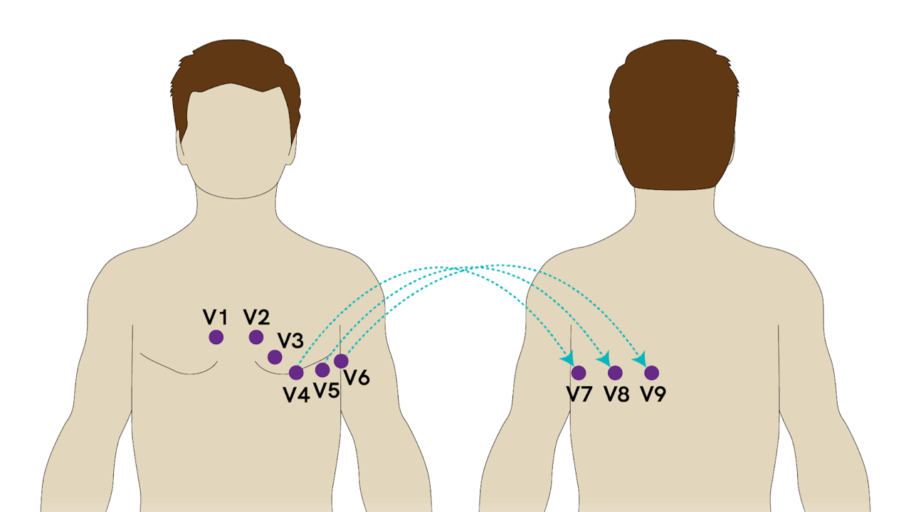
The posterior myocardium is not directly visualized on a standard 12 lead ECG, but reciprocal changes of STEMI in the anteroseptal leads (V1- V3) are seen (the posterior electrical activity is recorded from the anterior side of the heart)
If in V1- V3 you see
* ST segment depression
* Tall R wave
* Upright T waves
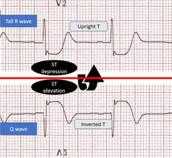
Consider posterior MI as a cause. You need to then obtain an ECG with posterior leads. If there is 0.5 mm elevation in any posterior lead this is diagnostic of posterior MI.
van Gorselen EO, Verheugt FW, Meursing BT, Oude Ophuis AJ. Posterior myocardial infarction: the dark side of the moon. Neth Heart J. 2007 Jan;15(1):16-21
Category: Trauma
Keywords: trauma, vasopressors, mass transfusion, uncertainty (PubMed Search)
Posted: 5/7/2023 by Robert Flint, MD
(Updated: 2/2/2026)
Click here to contact Robert Flint, MD
This extensive review looks at the literature surrounding vasopressors in trauma. Take away points are:
1. Most of the studies were done when the use of crystalloid was still being used as initial resuscitation fluid instead of blood.
2. Use of whole blood and mass hemorrhage protocols are not reflected in the literature regarding vasopressor use.
3. There are physiologic reasons vasopressors could be useful, particularly in head injured patients where we want increased mean arterial pressures.
4. European guidelines include vasopressor use whereas American ones do not.
5. Vasopressin and norepinephrine appear to be the vasopressors of choice if using a vasopressor in a trauma patient.
6. We need better studies looking at this topic
7. We need better studies looking at permissive hypotension in trauma now that our resuscitative strategy emphasizes mass hemorrhage protocol of blood, blood products, TXA and hemorrhage control.
8. As with all things in medicine, never say never.
Richards, Justin E. MD*; Harris, Tim MD†,‡; Dünser, Martin W. MD§; Bouzat, Pierre MD, PhD?; Gauss, Tobias MD¶. Vasopressors in Trauma: A Never Event?. Anesthesia & Analgesia 133(1):p 68-79, July 2021. | DOI: 10.1213/ANE.0000000000005552
Category: Pediatrics
Keywords: Pediatrics, infections, neonatal (PubMed Search)
Posted: 5/5/2023 by Rachel Wiltjer, DO
(Updated: 2/2/2026)
Click here to contact Rachel Wiltjer, DO
Neonatal rashes are common and, usually, benign. There are some skin findings, however, that require early recognition and treatment for best outcomes. One of these concerning etiologies is omphalitis, infection of the umbilical stump and surrounding tissues.
Features of omphalitis may include erythema and induration around the umbilicus, purulent drainage, and potentially systemic illness.
Risk factors include poor cord hygiene, premature or prolonged rupture of membranes, maternal infection, low birth weight, umbilical catheterization, and home birth.
Evaluation includes surface cultures from the site of infection as well as age-appropriate fever workup if patient is febrile. Consider ultrasound to evaluate for urachal anomalies as these can co-exist.
Management is IV antibiotics to cover S. aureus and gram negatives with surgical consultation if there are signs of necrotizing fasciitis or abscess. Some newer literature suggests that patients with omphalitis seen and treated in high-income countries may not be as sick as previously thought (as most data has been obtained in lower income countries where incidence is higher) and there has been a suggestion that there may be a role for oral antibiotics in well appearing, lower risk infants. This deserves further exploration but cannot yet be considered standard of care.
Other umbilical cord findings to consider (when it isn’t omphalitis): patent urachus, granuloma, local irritation, or partial cord separation
Kaplan RL. Omphalitis: Clinical Presentation and Approach to Evaluation and Management. Pediatr Emerg Care. 2023;39(3):188-189.
Category: Critical Care
Keywords: etomidate, intubation, critically ill, mortality (PubMed Search)
Posted: 5/2/2023 by Quincy Tran, MD, PhD
(Updated: 2/2/2026)
Click here to contact Quincy Tran, MD, PhD
As emergency physicians, we use etomidate to intubate patients most of the time, although there was controversy whether etomidate would suppress critically ill patients’ cortisol production. Whether etomidate was associated with mortality was controversial. A recent meta-analysis investigated the issue again.
Methods: meta-analysis of randomized trials using etomidate for intubation versus other agents. Outcome = mortality as defined by the authors. Mortality was defined from 24 hours to 30 days by study’s authors.
Results: 11 RCTs, including one new RCT in 2022
319 (1359, 23%) patients received etomidate died vs. 267 (1345, 20%) receiving other agents died; Risk Ratio 1.16, 95% CI 1.01-1.33, P = 0.03.
Etomidate was also associated with higher risk ratio for adrenal insufficiency, when compared with other control agents (147/695, 21% vs. 69/686, 10%, RR 2.01, 95% CI (1.59-2.56), P < 0.01.
Etomidate was also associated with higher risk ratio of mortality, when compared with ketamine, for mortality, as defined by each study’s author (273/1201, 23% vs. 226/1198. 19%. RR 1.18, 95% CI 1.02-1.37, P = 0.03).
Discussion:
The authors used fixed effects model, as they claimed that their meta-analysis had low heterogeneity (I2 =0%). However, fixed effects model should only be used when there is no difference among patient population. In this study, the outcome definitions were different, the patient populations were different (trauma, pre-hospital, ED, ICU). Therefore, random effects model should be used. Random effects models tend to yield larger 95% CI, thus, more likely yield non-statistically significant results.
The authors claimed a Number Needed To Treat (NNT) for etomidate of 31, so basically many ED patients would die, while most of patients being intubated by Anesthesiology, regarding settings, would not die, as anesthesiologists mostly use propofol.
Category: Cardiology
Keywords: POCUS, ACS, Regional Wal Motion Abnormality, Ultrasound (PubMed Search)
Posted: 5/1/2023 by Alexis Salerno Rubeling, MD
(Updated: 2/2/2026)
Click here to contact Alexis Salerno Rubeling, MD
In this study the researchers looked at patients presenting to the emergency department with high suspicion for ACS and explored if Regional Wall Motion Abnormality (RWMA) evaluation by EPs was associated with occlusion myocardial ischemia (OMI).
FOCUS identified RWMA in 87% of patients with coronary angiography proven OMI. With a sensitivity of 94%, specificity 35%, and overall accuracy of 78%.
The authors concluded that using FOCUS can have good utility when a patient is high risk for OMI and has an equivocal ekg. However, if RWMA is not present, physicians should still continue with work up such as cardiac catheterization.
To evaluate RWMA it is easiest to:
For more information check out this ACEPnow article: https://www.acepnow.com/article/detect-cardiac-regional-wall-motion-abnormalities-point-care-echocardiography/?singlepage=1
Bracey A, Massey L, Pellet AC, Thode HC, Holman TR, Singer AJ, McClure M, Secko MA. FOCUS amay detect wall motion abnormalities in patients with ACS, A retrospective study. Am J Emerg Med. 2023 Apr 2;69:17-22. doi: 10.1016/j.ajem.2023.03.056. Epub ahead of print. PMID: 37037160.
