Elderly patients in general have a lower baseline body temperature than younger patients. Consequently, it makes sense to redefine the definition of what constitutes a "fever" in the elderly. Rather than using the typical oral temperature cutoff of 38o C (100.4o F) for defining a fever, instead consider using 37.2o C (99o F). Redefining fever in this way increases the sensitivity for detecting bacterial infections from 40% to 83% while retaining an 89% specificity.
Saturday night palsy - radial nerve mononeuropathy due to improper arm positioning associated with inebriated sleep.
Physical examination - Wrist and finger drop.
Patients may have findings suggestive of ulnar nerve co-involvement (interossei testing) which may falsely lead the examiner to consider a more proximal location for the lesion such as the brachial plexus.
The finger drop caused by the radial nerve lesion places the hand at a mechanical disadvantage. Adjust for this by examining the hand on a flat surface (stretcher, counter top). With the fingers now supported in extension at the MCP joint (no longer "dropped"), the interossei can now be tested in isolation and will be normal.
EYE OPENING
4 = spontaneous
3 = to voice
2 = to pain
1 = none
VERBAL RESPONSE
5 = orientated
4 = confused
3 = inappropriate
2 = incomprehensible
1 = none
MOTOR RESPONSE
6 = obeys command
5 = localizes pain
4 = withdraw to pain
3 = decorticate
2 = decerebrate
1 = none
Spontaneous Bacterial Peritonitis
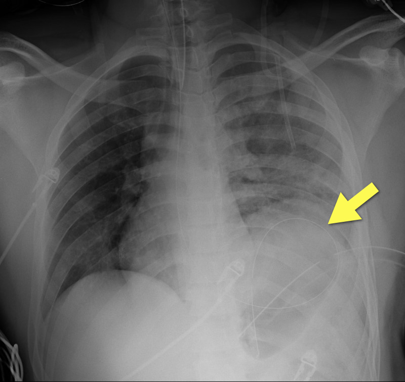
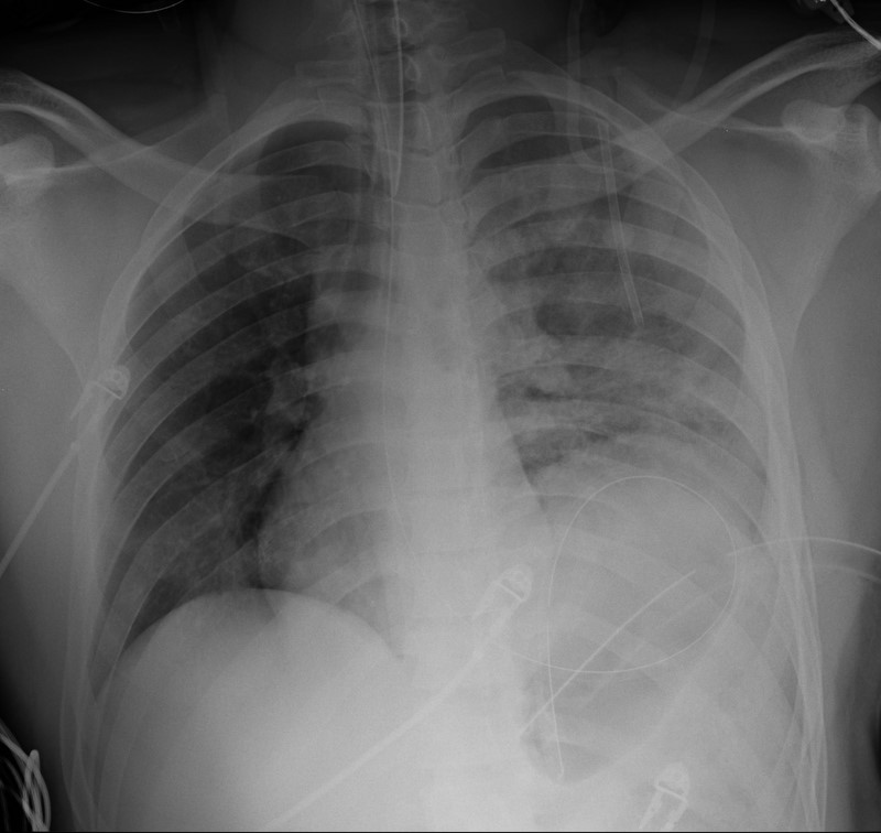
19 year-old male s/p high-speed MVC with hypotension and diminished breath sounds on left. Diagnosis?
A patient presents to the ED in pulmonary edema, hypotensive, and has JVD. There's a new systolic murmur. The patient had an acute MI 7-10 days ago and had appropriate treatment and uncomplicated course, then discharge. What's the diagnosis and what do you do?
Step 1: Sign out immediately.
Step 2: If it's not time to sign out (just kidding about step 1), listen carefully to the murmur. If it's heard best at the lower sternal border, it's probably a ruptured papillary muscle with acute MR. If it's a "machinery" type murmur heard throughout the precordium loudly, it's probably an acute VSD.
Step 3: VSD patient is likely to die, but with either one, you've got to move quickly. IMMEDIATELY call cardiology AND cardiac surgery. The patient is in need of a balloon pump and OR.
All you can do is buy time until the patient goes upstairs....pressors for BP, IV NTG as tolerated for preload reduction, and be judicious with diuretics. Vasodilators might help unload the heart also. This patient may end up on 2-3 drips, and make sure ALL meds are titrateable. And just keep your fingers crossed!
Posterolateral Corner Injuries
The posterolateral corner “PLC” of the knee is becoming increasingly recognized as an extremely important structure to maintain the stability of the knee joint.
PLC injuries occur with hyperextension, varus load and tibial external rotation. So the most common mechanism is a posterolaterally directed blow to the anteromedial tibia when the knee is hyperextended. PLC injuries are commonly associated with injury to other ligaments (ACL, PCL, LCL) and occur in isolation in <5% of cases. If suspected make sure to check for other ligamentous injuries.
Since this injury can be missed and is associated with significant disability it is important to test for it. This YouTube video, http://youtu.be/bnXaTdvZZ6o, demonstrates several examination techniques that can identify the injury.
Radial and femoral arteries are common sites for arterial-line placement, but are not without complications (e.g., Radial artery: malfunction with positioning and Femoral artery: contamination and infection); an alternative site to consider is the axillary artery.
The axillary artery's superficial location and large size make it a desirable choice for cannulation.
The "anatomical-landmark" and "palpation" methods have been the traditional techniques of axillary arterial cannulation, however these methods may be difficult for to a variety of reasons (e.g., obesity, anasarca, arterial disease, etc.)
Ultrasound allows visualization of the axillary artery and avoids unintended injury to structures in close proximity (e.g., brachial plexus, pleura, axillary vein, etc.); please see figures 1 and 2 in the referenced Sandhu article and http://www.youtube.com/watch?v=Z31YiyV7cNQ.
A recent study (Killu, 2011) found that ultrasound increases success rates when compared to the traditional landmark approach.
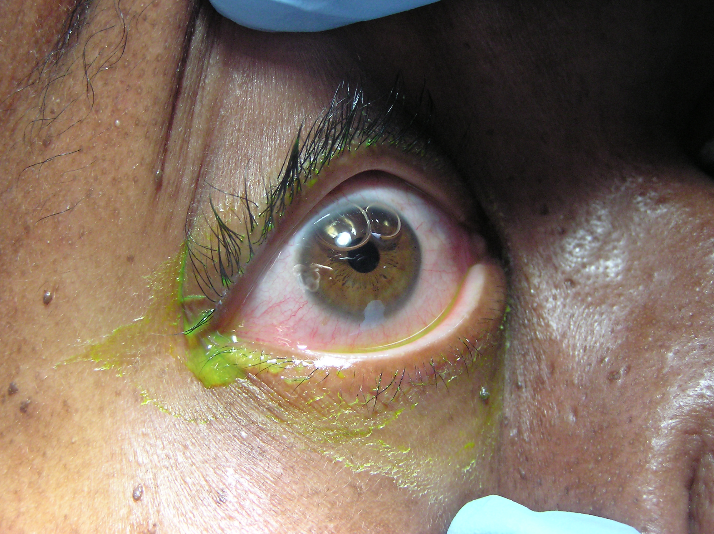
A 50 year-old patient presents after a self-inflicted eye injury. The patient had taken some type of needle and inserted it into their eye.
What is the diagnosis and what complications might result?
SVT is rarely, if ever, the presenting rhythm associated with an acute MI. As a result, physicians should not feel compelled to send troponin levels and perform rule-outs purely based on an SVT presentation. Instead, the decision to rule out a patient presenting with SVT should be based on whether there is a constellation of other concerning symptoms, exclusive of the SVT (e.g. if the patient presented with chest pressure radiating down the arm and diaphoresis, in addition to the SVT).
Two recent studies confirmed that routine troponin testing in patients with SVT is extremely low-yield, and instead often produces false-positive troponin results that lead to unnecessary admissions and workups. In other words, mild troponin elevations may occur in SVT but they do not correlate with true ACS.
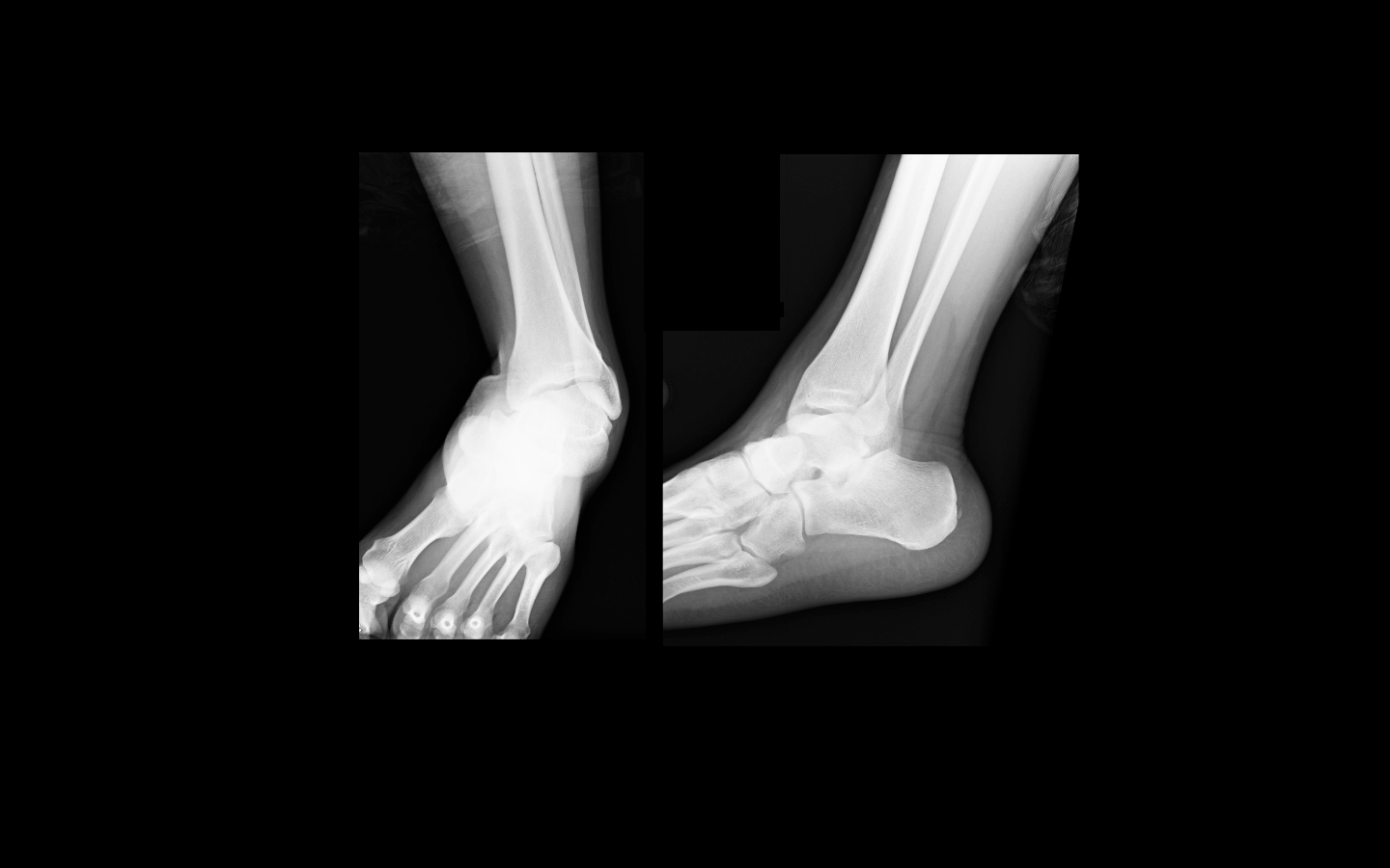
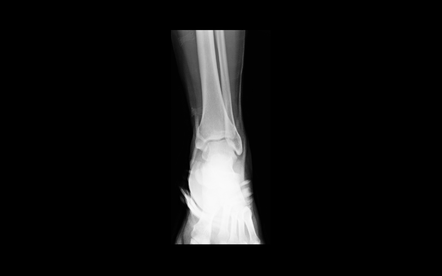
Evaluation of circulatory status is the most important aspect of post reduction care.
Look for hard findings such as cool/cold lower extremity, diminished or absent pulses, pale or dusky skin, paralysis, etc.
However, the absence of these findings should not lull the clinician into a false sense of security. The degree of initial joint deformity, presence of full bounding pulses and warm skin over the dorsum of the foot can all be present in the setting of vascular injury.
The next step will be to perform an ABI (ankle-brachial index).
In one small study, no patient with an ABI greater than or equal to 0.9 had a vascular injury.
Patients with a reassuring physical exam and ABIs should be admitted for vascular checks without further imaging.
Patients with a reassuring physical examination but with an abnormal ABI should have an imaging study obtained (arteriogram/CT angiogram).
Patients with hard findings of a vascular injury should have an emergent vascular surgery consultation.
Every so often a patient arrives in PSVT with their only intravenous access being through a hemodialysis port.
Initial dose of adenosine should be reduced to 3 mg if administered through a central line. Remember a central line delivers the adenosine right where you need it. This recommendation is supported by the 2010 ACLS guidelines. Second and third doses should be 6 mg (instead of 12 mg).
Cases of prolonged bradycardia and severe side effects have been reported after full-dose adenosine through a central line. Other situations to consider lower doses include patients currently receiving carbamazepine or dipyridamole or in those with a transplanted heart.
Fungal Sepsis in the Critically Ill
Approximately 7-10% of cases of ACS are not related to atherosclerotic coronary disease. Some other causes of ACS include the following:
trauma
vasculitis
congenital abnormalities
emboli (e.g. bacterial)
thoracic aortic dissection
infectious diseases
DIC, TTP
These conditions can produce ST-segment changes that resemble those of true STEMI or non-STEMI, and therefore some of these patients are diagnosed retrospectively after a negative catheterization.
Sugar Tong Splint
The sugar tong splint is ideal for splinting fractures of the radius, ulna, or wrist. It prevents flexion and extension at the wrist, limits flexion and extension at the elbow, and prevents supination and pronation. A posterior long arm splint does not prevent supinaton and pronation, therefore, it is of limited use for radius and ulna fractures.
The traditional sugar tong can be difficult to put on a patient without an assistant as it is often hard to hold the splint in position as you begin to ace wrap it. A variation on the sugar tong, the reverse sugar tong, prevents this frustration. The splinting material is cut so that a small piece suspends the splint from the web space between the thumb and index finger. The open ends at the elbow are also easily folded under each other, preventing any bulky splint material from extending out.
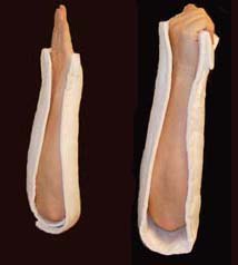
The reverse sugar tong is on the left, the original sugar tong on the right.
Check out this video showing how to place a reverse sugar tong splint.
http://www.youtube.com/watch?v=r-RHdttOMf0