Extracorporeal Treatment Strategies for Acute Methanol Poisoning (When to Dialyze)
Methanol toxicity is classically included in the differential for the intoxicated patient presenting to the ED. Add a negative EtOH level, anion/osmolar gap, blindness and you have yourself a slam dunk diagnosis. The goal is to stop the liver from metabolizing methanol to formic acid. Outside of fomepizole (or old school ethanol therapy), dialysis is often discussed, but when should you actually get the nephrologist on the phone?
This month the Extracorporeal Treatments in Poisoning Workgroup released a systematic review and consensus statement to help clinicians decide when to pull the HD trigger. Their suggestions are below.
When to start HD:
Which Modality: Intermittent HD (IHD) should be used over continuous renal replacement therapies (CRRT), as you can clear the toxin faster with higher HD flows.
When to stop HD: Extracorporeal treatment can be terminated when the methanol concentration is less than 200 mg/L or 6.2 mmol/L and a clinical improvement is observed.
Bottom Line: Consider early hemodialysis in most patients presenting with methanol toxicity. Clinical exam and routine lab testing will likely provide enough information to determine the need for IHD, but specific methanol levels can be helpful to guide adjunctive treatment options.
Reference
Roberts DM, Yates C, Megarbane B, et al. Recommendations for the Role of Extracorporeal Treatments in the Management of Acute Methanol Poisoning: A Systematic Review and Consensus Statement. Crit Care Med. 2015;43(2):461-472.
Follow me on Twitter @JohnGreenwoodMD
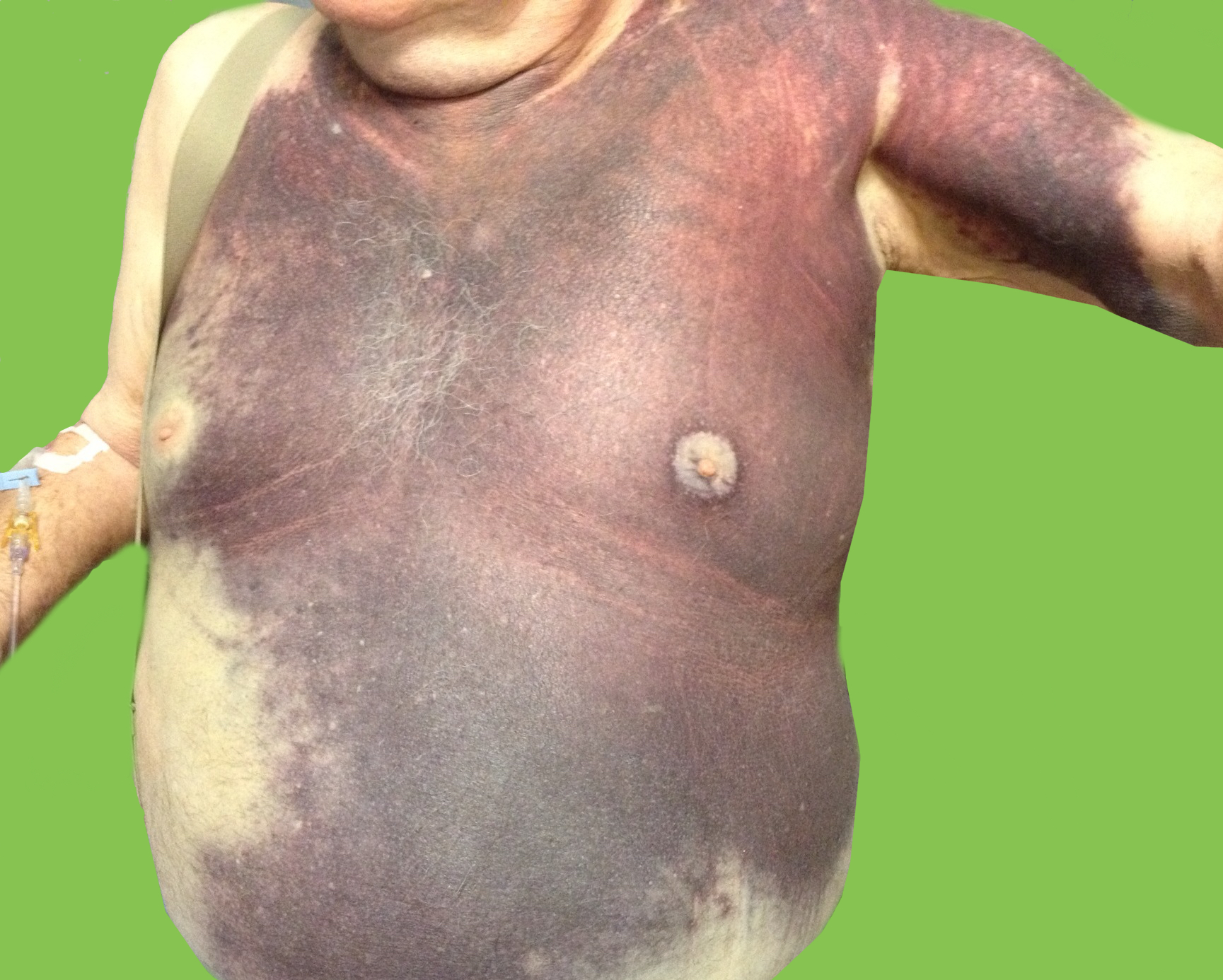
Elderly male presents with the skin findings below. He is also on a medication for atrial fibrillation. What's the diagnosis?
Posterior Myocardial Infarctions (PMI)
- Posterior myocardial infarctions (PMI) are different than typical ST-elevation MI; the ECG findings include: septal & anterior ST-segment depression, dominant tall/broad R waves, and upright T waves.
- In a study among 117,739 subjects with STEMI, 824 with PMI were more likely to present with cardiac arrest, cardiogenic shock, and congestive heart failure.
- The median time from arrival ECG to revascularization with PCI was longer among subjects with PMI.
- The median time from arrival ECG to systemic thrombolysis was also longer among subjects with a PMI.
- Increased awareness and recognition of PMI is needed to improve reperfusion times among this subpopulation with STEMI.
Overuse injury
Seen in runners, military recruits (marching), ballet dancers and in jumping sports (heavy landing).
Insidious onset of heel pain, that is worse with jumping then running then later with simple weight bearing.
Tenderness to palpation posteriorly (medially or laterally), and squeezing bilateral posterior calcaneus.
Testing:
XR: May not be positive for 2 to 4 weeks. Sclerotic appearance (vertically oriented) posterior calcaneus.
MRI: high signal T2 at fracture site.
DDx: plantar fasciitis.
Treatment: Reduction of activity if Sxs mild, for severe pain start a trial of non weight-bearing (boot or splint with crutches).
Stretching of calf, achilles, plantar fascia.
As noted previously (UMEM Pearl of 1/7/2015), tuberculosis (TB) is a major infectious disease that occurs worldwide. Strains of tuberculosis can be resistant to one or more anti-tuberculosis medications. TB strains resistant to at least one medication have been found in all surveyed countries.
What is multidrug-resistant tuberculosis (MDR TB)?
Treatment of MDR TB
Bottom line:
As noted previously, in your emergency department have a high index of suspicion for TB and MDR TB in patients with an appropriate risk profile.
60 year-old male presents with 6 months of weight loss,epistaxis, and increased headache when bending over. What's the diagnosis?
Of pediatric patients who have anteroposterior (AP) pelvic xrays (XR), there is a 4.6% rate of pelvic fracture or dislocation, compared to 10% in adults.
This study is a sub analysis of a prospective observational cohort of children with blunt torso trauma conducted by PECARN. 7808 patients had pelvic imaging, with 65% of them having an AP XR. The XR sensitivity ranged from 64-82% (based on age groups) for detecting fractures. All but one patient with a pelvic fracture not detected on XR had a CT scan. The CT scan detected all but 2 fractures both of which were picked up later as healing fractures on repeat pelvic XR. Some of the patients who had a missed fracture on XR were hemodynamically unstable or wound up requiring operative intervention.
The authors support the following algorithm:
-With hemodynamically unstability children, obtain a pelvic XR
-For hemodynamically stable children when the physician is planning to get a CT, there is no indication for XR
Bottom line: Consider using AP pelvic radiographs in the hemodynamically stable patient with a high suspicion for fracture or dislocation who are not undergoing CT.
Left Ventricular Hypertrophy & Arrhythmias: Any Association?
Associations between left ventricular hypertrophy (LVH) and both supraventricular (SVT)/ventricular arrhythmias (VT/VF) have previously been reported.
A recent review & meta-analysis of 10 studies (27,141 patients) revealed the following:
- Incidence of SVT was 11% with LVH compared to 1% without (p <0.001)
- LVH patients had 3.4-fold greater odds of developing SVT
- Incidence of VT/VF was 5.5% with LVH compared to 1.2% without (p <0.001)
- LVH patients has 2.8 greater odds of developing VT/VF
The reason for increased arrhythmogenicity in LVH is not clearly understood.
A consistently observed abnormality in LVH is non-uniform propagation of the action potential throughout the myocardium, which sets the stage for arrhythmias based on early or delayed afterdepolarizations.
Given the heterogeneity in this meta-analysis further research between LVH & sustained arrhythmias is needed to infer true causality.
We often think of Plantar Fascitis as the cause of heel pain but there are a lot of other causes. Some of those include:
Hydroxocobalamin is an effective cyanide antidote when administered intravenously. Although intraosseous (IO) access is often used in critically ill patients with difficult or delayed IV access, the efficacy of IO administration has not been investigated until recently.
In a recent randomized animal study, acute cyanide toxicity was induced in two groups of swine where 150 mg/kg Hydroxocobalamin was administered via IV vs. IO. The survival rate, reversal of hypotension, and laboratory results were similar between the IV and IO group.
The finding of this study suggest that IO administration of Hydroxocobalamin is as efficacious as IV administration and its administration in acute cyanide toxicity should not be delayed due to lack of IV access when IO access is available.
Is progesterone yet another disappointing neuroprotectant?
Traumatic brain injury (TBI) affects more than 1.7 million persons in the U.S. annually. The incidence of TBI is increasing globally, especially in developing countries. Despite improvement in trauma systems and critical care, the morbidity and mortality associated with severe TBI remain high.
Progesterone has been shown in preclinical and phase 2 randomized clinical trials to have pluripotent neuroprotective properties and improve mortality in TBI.
Two multicenter phase 3 trials were recently completed and published in the December 25th issue of the New England Journal of Medicine. However, their results were disappointing.
Both studies used the Glasgow Outcome Scale (GOS) or Extended Glasgow Outcome Scale (GOS-E) at 6 months as their primary outcome. The GOS and GOS-E capture the degree of recovery from brain injury in terms of disability, stratified into levels by death, vegetative state, severe disability, moderate disability, and good recovery.
Progesterone was not found to have any benefit in functional outcome at 6 months.
Both of these studies were well designed and conducted. However, they were based on small effect sizes of the phase 2 trials. In addition, they had very favorable outcome rates in the placebo group, thereby making it hard to demonstrate a benefit by their sample sizes.
There has been a long history of failed neuroprotectant trials likely due to the complex and variable injury mechanisms involved in TBI. The currently available outcome measures are also insensitive to the targeted mechanistic endpoints. More research is needed to understand not only potential therapies but also how to select appropriate patients for these therapies.
Take Home Point: Progesterone does not have any clear benefit in TBI at this time.
Diaphragm weakness and its significance
There are several ways to monitor diaphragm strength and function
Clinical Relevance
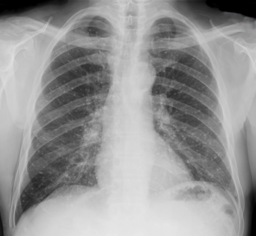
60 year-old male presents with rhinorrhea, body aches, and dry cough for two days. He recently moved here from Kentucky. What’s the diagnosis (please note that there are several differentials based on CXR)?
APACHE-HF Scoring System
The Acute Physiology and Chronic Health Evaluation (APACHE) scoring system was established in the 1980's to predict critically ill patient prognosis (APACHE II, III, and IV have been published in last two decades).
The APACHE II scoring system involves combining 3 separate scores (acute physiology score, chronic health score, and age), which can be cumbersome to apply & thus is not often utilized in the emergency department (modified APACHE II doesn't include chronic health score & is less taxing).
No unique scoring system for acute heart failure (AHF) has been analyzed until present; the APACHE-HF score includes 8 criteria: mean arterial pressure (MAP), pulse, sodium, potassium, hematocrit, creatinine, age, and glasgow coma score (GCS).
AHF in-hospital mortality data was analyzed and compared using APACHE II, modified APACHE II, and APACHE- HF scores and the predictive value of the APACHE-HF score was found to be optimal when compared to the others.
Mechanism: Direct blow to anterior thigh (Football/basketball) or from a ball traveling at a high rate of speed (field hockey/lacrosse).
Exam: focal tenderness and edema. Pain may be severe and worse with active contraction and passive stretch. Hematoma may already be present. Amount of passive knee flexion at 24 hours can be a clue to the severity of the hematoma.
Treatment: Crutches if pain with weight-bearing. Ice. Immobilization in 120 degrees of flexion immediately after the injury for the first 24 hours may be beneficial.
-Bandage entire lower limb. Provide crutches and pain medication. Soft tissue therapy is contraindicated for the first 48 hours and when instituted must be gentle and cause no pain. Risk of re-bleeding is greatest in first 7 to 10 days.
http://fce-study.netdna-ssl.com/2/images/upload-flashcards/75/20/63/5752063_m.jpg
Application to Clinical Practice
WBI can facilitate removal of select toxicants from the gastrointestinal tract in some patients, but there is no convincing evidence from clinical studies that it improves the outcome of poisoned patients.
Tuberculosis (TB) is a major infectious disease that occurs worldwide.
Fortunately, public health and clinical measures have had some success.
Bottom line:
"PQRST" - Capnography in Cardiac Arrest